For Providers -- Atlas of Human Anatomy
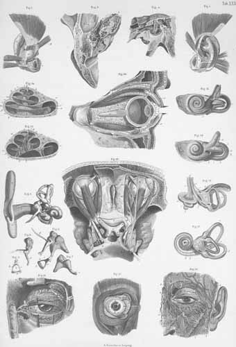
Plate 31: Visual and hearing apparatus.
Translated by: Ronald A. Bergman, PhD and Adel K. Afifi, MD, MS
Peer Review Status: Internally Peer Reviewed
- The anterior surface of the (left) outer
ear cartilage and outer ear muscles.
- Posterior surface of the (left) outer
temporal bone with the external muscles.
- The interior (tympanic cavity, cochlea,
vestibule) of the left ear, open from above.
- The left tympanic cavity, (after removal
of the inner wall of the tympanic membrane), with auditory ossicles.
- The parts of the right auditory organ connected.
- Hammer, Incus (ambos), and Stapes.
- Incus (of left ear).
- Hammer (malleus), of left ear.
- Stapes, of left ear.
- Bones of the labyrinth (of left ear)
outer surface, unopened.
- The bony labyrinth, of the left ear, opened.
- The bones and membraneous labyrinth (of
the left ear) with its nerves.
- Opened membranous labyrinth (of the left
ear).
- Bony cochlea (of left ear), opened
from the side.
- The bony cochlea (of left ear), vertical
middle section.
- The left eye of the dissected face,
with its muscles and nerves.
- The outer and inner muscles of the (right)
eye, and exposure of the internal stratum of musculus orbicularis.
- The tear and eyelid glands of the
left eye.
- Both orbital cavities, opened from above,
with their muscles and nerves.
- The right ocular bulb in the orbit,
cut through the middle, opened from the lateral side.
|
 |
Title Page