For Providers -- Atlas of Human Anatomy

Both orbital cavities, opened from above, with their muscles and nerves.

Translated by: Ronald A. Bergman, PhD and Adel K. Afifi, MD, MS
Peer Review Status: Internally Peer Reviewed


Both orbital cavities, opened from above, with their muscles and nerves

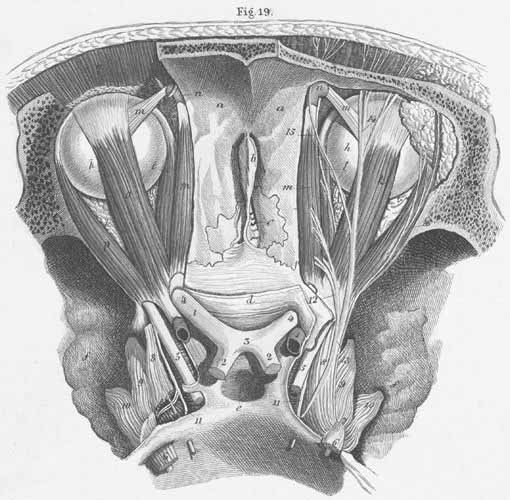
a) Inner surface of frontal bone.
b) Crista galli.
c) Lamina cribrosa.
d) Body of sphenoid bone.
e) Sella turcica (fossa for hypophysis [pituitary gland]).
f) Greater wing of the sphenoid bone.
g) Outer wall of the orbit.
h) Ocular bulb.
i) Lacrymal gland.
k) m. Superior rectus oculi.
l) m. Medial rectus oculi.
m) m. Superior oblique oculi.
n) Trochlea of m. superior oblique.
o) m. Levator palpebral superioris.
p) m. Lateral rectus oculi.
q) m. Frontal.

  1. ophthalmic artery.
  2. optic tract.
  3. optic chiasm.
  4. optic nerve.
  5. oculomotor nerve.
  6. trigeminal nerve.
  7. semilunar ganglion (s. trigeminal or Gasserian ganglion).
  8. trigeminal nerve, ophthalmic division.
  9. trigeminal nerve, maxillary division.
  10. trigeminal nerve, mandibular division.
  11. abducens nerve.
  12. trochlear nerve.
  13. ophthalmic nerve, frontal branch.
  14. supraorbital nerve.
  15. Lacrymal nerve.

Plate Index | Title Page