For Providers -- Atlas of Human Anatomy
Translated by: Ronald A. Bergman, PhD and Adel K. Afifi, MD, MS
Peer Review Status: Internally Peer Reviewed
Magnified View (via Quicktime VR)
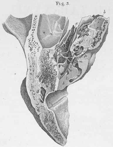
a) Temporal bone, pars squamosa.
b) Temporal bone, pars petrosa.
c) Tympanic cavity.
d) Tympanic membrane (inner surface).
e) Head of malleus.
f) Incus (s. corpus incudus).
g) Incus, short process.
h) Incus, long process.
i) Head of stapes.
k) Stapes, anterior crus.
l) Stapes, posterior crus.
m) Footplate (s. basis) [in oval window].
n) m. Tensor tympani.
o) m. Stapedius.
p) Internal auditory meatus.
q) Cochlea.
r) Spiral lamina.
s) Modiolus.
t) Scala tympani.
u) Scala vestibuli.
v) Atrium or vestibule.
w) Posterior semicircular canal.
x) Eustachian tube (auditory tube).
y) Mastoid air cells.
z) Facial canal (s. canalis Fallopii) [for the facial nerve].