For Providers -- Atlas of Human Anatomy
Translated by: Ronald A. Bergman, PhD and Adel K. Afifi, MD, MS
Peer Review Status: Internally Peer Reviewed
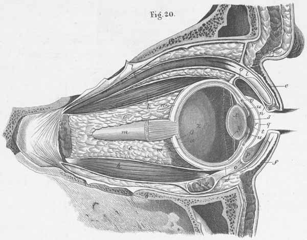
a) Frontal bone.
b) Maxillary bone.
c) Fat in orbit.
d) m. Frontal.
e) Superior eyelid.
f) Inferior eyelid.
g) m. Inferior oblique oculi.
h) m. Inferior rectus oculi.
i) m. Lateral rectus oculi.
k) m. Superior rectus oculi.
l) m. Levator palpebral superioris.
m) Optic nerve.
n) Conjunctiva of eyelid.
o) Transition of eyelid and skin in the
p) sclera conjunctiva (ocular bulb).
q) Corneal conjunctiva.
r) Cornea.
s) Decemet’s membrane (lamina limitans posterior cornea).
t) Anterior ocular cavity, with aqueous humor.
u) Posterior ocular cavity, with aqueous humor.
v) Iridial sinus venosus.
w) Sclera, tunica.
x) Crystalline lens (capsule and Petit’s canal).
y) Ciliary body.
z) Vitreous body.
a) Retina, tunica.
b) Choroid, tunica.