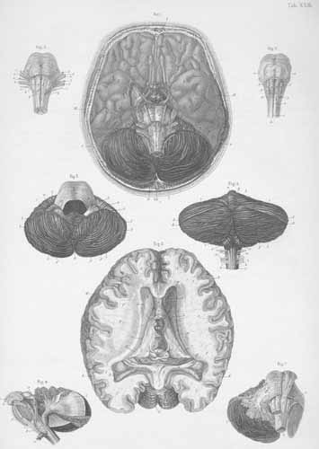
- Base of the brain and location of cranial nerves.
- Inner surface of the brain, seen from above, exposing both lateral ventricles and the third ventricle. The corpus callosum, septum pellucidum and fornix seen in front of 3rd ventricle. The foramen of Monro is cut across and passes posteriorly.
- Inferior surface of the cerebellum, after removal of the medulla oblongata.
- Cerebellum and proximal end of the spinal cord, seen from below.
- Pons and medulla oblongata.
- Pons (Varoli) and medulla oblongata with the internal fibers of each.
- Pons (with its transverse and longitudinal fibers), medulla oblongata and right hemisphere of the cerebellum.
- The long fiber path of the medulla oblongata, which course from the cerebellum, corpora quadrigemina to the pons.