For Providers -- Atlas of Human Anatomy
Translated by: Ronald A. Bergman, PhD and Adel K. Afifi, MD, MS
Peer Review Status: Internally Peer Reviewed
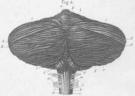
A. Hemisphere of cerebellum.
B. Superior vermis of cerebellum.
C. Inferior vermis of cerebellum.
D. Spinal cord.
E. Great horizontal sulcus (s. Reilii).
a) Central lobule, monticulus cerebelli (central part of superior vermis).
b) Culmen, folium cacuminis.
c) Quadrangular lobule, [lobulus quadrangularis (s. superior anterior)].
d) Tonsil.
e) Biventer lobule, (Lobulus ansiformis).
f) Lobulus gracilis (s. tener).
g) Inferior semilunar lobule, (s. inferior posterior).
h) Pyramid of vermis.
i) Lateral funiculus (spinal cord).
k) Fasciculus cuneatus (spinal cord).
l) Fasciculus gracilis (spinal cord).
m) posterior (dorsal) roots of spinal nerves.