For Providers -- Atlas of Human Anatomy
The thoracic and abdominal cavities are opened from the front and important
lymph glands and their vessels are shown. Viscera of the left side are removed.
Translated by: Ronald A. Bergman, PhD and Adel K. Afifi, MD, MS
Peer Review Status: Internally Peer Reviewed
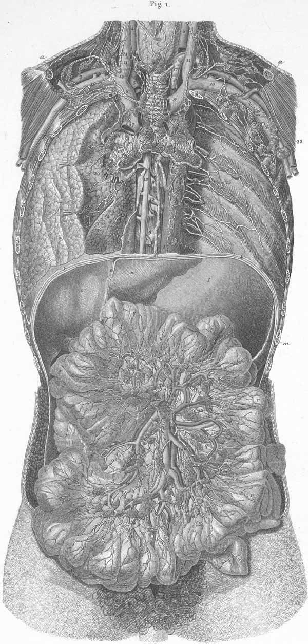
a) Clavicle.
b) First rib.
c) Thyroid gland.
d) Trachea.
e) Right bronchus.
f) Left bronchus.
g) Thoracic vertebral column.
h) Right lung.
i) Posterior mediastinum.
k) Diaphragm.
l) Stomach, body (s. ventriculus).
m) Spleen (s. lien).
n) Liver, left lobe.
o) Liver, right lobe.
p) Ascending colon (s. right colon).
q) Mesentery (of small intestine).
r) Jejunum and Ileum.
s) Gallbladder (s. vesica fella).
t) Suspensory ligament of liver.
- aortic arch.
- descending thoracic aorta.
- subclavian artery.
- common carotid artery.
- innominate artery (s. brachiocephalic artery).
- intercostal artery and veins.
- superior vena cava.
- innominate vein (s. right common jugular vein).
- left innominate vein (s. transversa).
- subclavian vein.
- internal jugular vein.
- azygos vein.
- hemiazygos vein.
- superior mesenteric artery (s. mesaraica superior).
- superior mesenteric vein (s. mesaraica magna).
- jejunal and ileal arteries and veins.
- thoracic duct.
- right common lymphatic trunk (s. minor).
- bronchial nodes.
- pulmonary nodes.
- deep jugular nodes.
- axillary nodes.
- intercostals nodes.
- mesenteric plexus with mesenteric nodes (s. mesaraicae).
Plate Index | Title Page