For Providers -- Atlas of Human Anatomy
Translated by: Ronald A. Bergman, PhD and Adel K. Afifi, MD, MS
Peer Review Status: Internally Peer Reviewed
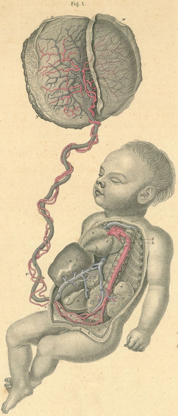
a) Right ventricle.
b) Left ventricle.
c) Left atrium.
d) Right pulmonary artery.
e) Aortic arch.
f) Pulmonary artery.
g) Left pulmonary artery.
h) Left pulmonary vein.
i) Ductus arteriosus.
k) Junction, ductus arteriosus and thoracic aorta.
l) Superior vena cava.
m) Left subclavian vein.
n) Common iliac artery.
o) External iliac vein.
p) Superior gluteal artery.
q) Internal iliac artery.
r) Umbilical artery.
s) Umbilical vein.
t) Urinary bladder and superior vesical artery.
u) Urachus.
v) Fetal blood vessels.
w) Amnion.
x) Chorion.
y) Maternal surface of placenta.
z) Liver, left lobe.
a) Liver, right lobe.
b) Gallbladder.
g) Umbilical vein.
d) Portal vein.
e) Ductus venosus.
z) Inferior vena cava.
h) Left hepatic vein.
q) Inferior vena cava.
l) Spleen.
m) Kidney, left.
n) Suprarenal (adrenal) gland.