For Providers -- Atlas of Human Anatomy
Translated by: Ronald A. Bergman, PhD and Adel K. Afifi, MD, MS
Peer Review Status: Internally Peer Reviewed
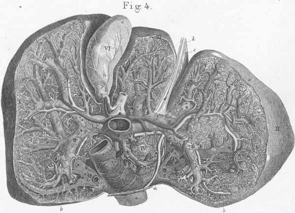
a) Anterior liver surface.
b) Posterior liver surface.
c) Suspensory ligament of liver.
d) Ligament teres hepatis (in umbilical fossa).
e) Inferior vena cava.
f) Fossa of ductus venosus (Aranti).
g) Portal vein.
h) Hepatic artery.
i) Ductus choledochus (s. bile duct).
k) Cystic duct.
l) Hepatic duct.
m) Ductus venosus (Arantii).
n) Artery to the gallbladder.
o) Gallbladder, fundus.
p) Gallbladder, neck.
q) Hepatic vein.