For Providers -- Atlas of Human Anatomy
The abdominal viscera seen after removal of the mesentery, as well as the
lungs, from the front.
Translated by: Ronald A. Bergman, PhD and Adel K. Afifi, MD, MS
Peer Review Status: Internally Peer Reviewed
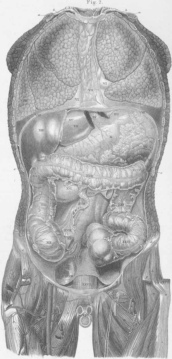
a) Clavicle.
b) First rib.
c) Eleventh rib.
d) Iliac crest (spine).
e) m. Psoas major.
f) m. Internal iliac.
g) m. Rectus femoris.
h) m. Gluteus medius.
i) m. Vastus lateralis.
k) m. Obturator externus.
l) Obturator membrane.
m) m. Adductor magnus.
n) m. Adductor brevis.
o) m. Adductor longus.
p) m. Gracilis.
q) m. Pectineus.
r) m. Tensor fasciae latae.
s) m. Sartorius.
t) m. Vastus intermedius.
u) Neck of the femoral bone.
v) Greater trochanter.
- femoral artery.
- femoral vein.
- superficial epigastric artery and vein.
- deep femoral artery and vein.
- lateral circumflex femoral artery and vein.
- Obturator nerve.
- Right lung, superior lobe.
- Right lung, middle lobe.
- Right lung, inferior lobe.
- Left lung, superior lobe.
- Left lung, inferior lobe.
- Pleura.
- Anterior mediastinum.
- Diaphragm.
- Esophagus.
- Fundus of stomach.
- Spleen (s. lien).
- Left lobe of liver.
- Right lobe of liver.
- Gallbladder (s. vesica fella).
- Suspensory ligament of liver.
- Duodenum.
- Jejunum.
- Mesentery.
- Cecum.
- Vermiform process (s. appendix).
- Transverse colon.
- Right colic flexure.
- Transverse colon.
- Left colic flexure.
- Descending colon.
- Sigmoid colon (s. iliac flexure).
- Rectum.
- Peritoneum.
- Ileum.
- Penis.
Plate Index | Title Page