For Providers -- Atlas of Human Anatomy

The deep nerves of the face (the left half). The zygomatic arch and the mandible are removed and the temporal fossa is opened.

Translated by: Ronald A. Bergman, PhD and Adel K. Afifi, MD, MS
Peer Review Status: Internally Peer Reviewed


The deep nerves of the face (the left half)

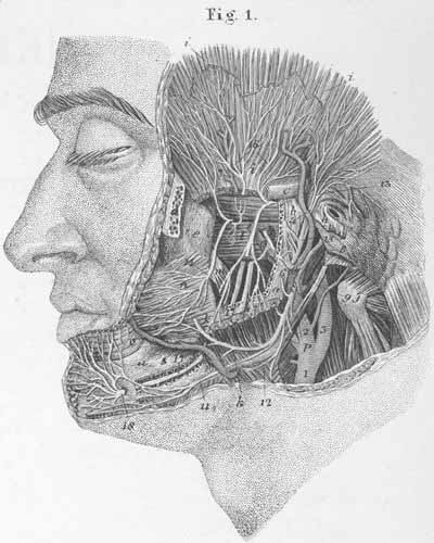
a) Mandible (s. maxilla inferior).
b) Temporomandibular joint.
c) Root of the zygomatic arch.
d) Zygomatic bone.
e) Maxillary bone.
f) Atlas, transverse process.
g) Styloid process.
h) External auditory meatus.
i) m. Temporalis.
k) m. Masseter.
l) m. Lateral pterygoid.
m) m. Medial pterygoid .
n) m. Buccinator.
o) m. Orbicularis oris.
p) m. Digastric, posterior belly.

  1. Common carotid artery.
  2. External carotid artery.
  3. Internal carotid artery.
  4. Temporalis artery.
  5. Facial artery.
  6. Maxillary artery.
  7. Deep temporal artery.
  8. Facial nerve.
  9. Facial nerve, superior (temporo-facial) ramus.
  10. Facial nerve, inferior (cervico-facial) ramus.
  11. Facial nerve, mandibular ramus.
  12. Superior cutaneous nerve of neck.
  13. Anterior auricular nerve.
  14. Buccinator nerve.
  15. Masseteric nerve.
  16. Deep temporal nerve.
  17. Inferior alveolar nerve
  18. Mental nerve.

Plate Index | Title Page