For Providers -- Atlas of Human Anatomy
Translated by: Ronald A. Bergman, PhD and Adel K. Afifi, MD, MS
Peer Review Status: Internally Peer Reviewed
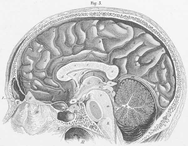
I. Frontal bone (with frontal sinus).
II. Crista galli (of ethmoidal bone).
III. Perpendicular lamina of the ethmoid bone.
IV. Body of the ethmoid bone.
V. Back of the sella turcica (posterior clinoid process).
VI. Sella turcica.
VII. Sphenoid sinus.
VIII. Basilar part of the occipital bone (with the fossa for medulla oblongata).
IX Occipital part of the occipital bone.
X. Vomer.
XI. Pharynx.
XII. Tentorium cerebelli (with confulence of sinuses and opened great cerebral
vein of Galen).
A. Anterior (Frontal) cerebral lobe [OBS].
B. Middle (Parietal) cerebral lobe [OBS].
C. Posterior (Parietal) cerebral lobe[OBS].
E. Medulla oblongata.
a) gyri.
b) sulci (furrow between gyri).
c) corpus callosum (body).
d) genu of corpus callosum.
e) corpus callosum, splenium.
f) septum pellucidum.
g) fornix (body).
h) fornix column.
i) Foramen of Munro.
k) Thalamus (optic thalamus).
l) anterior commissure.
m)
n) posterior commissure.
o) pineal gland.
p) stalk of pineal gland (s. crus glandulae pinealis).
q) corpora quadrigemina.
r) pons Varoli.
s) aqueduct of Sylvius.
t) tuber cinereum.
u) infundibulum.
v) pituitary gland (s. hypophysis).
w) optic chiasm.
x) optic nerve.
y) fourth ventricle.
z) mamillary body (s. candiacans).
a) anterior cerebellar valvule.
b) anterior cerebral artery.