For Providers -- Atlas of Human Anatomy
Translated by: Ronald A. Bergman, PhD and Adel K. Afifi, MD, MS
Peer
Review Status: Internally Peer Reviewed
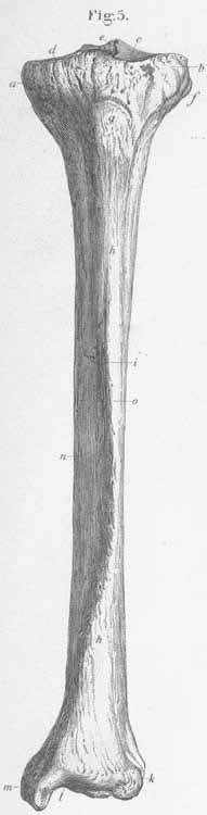
a) Medial condyle.
b) Lateral condyle.
c) Medial condylar cavity.
d) Lateral condylar cavity.
e) Intercondylar eminence (insertion for cruciate ligaments).
f) Superfical peroneal glenoid (socket) (for the fibular head).
g) Tibial spine s tuberosity (insertion site for patellar ligament).
h) Tibial body.
i) Tibial crest (sharp anterior edge).
k) Fibular notch (for the lower end of the fibula).
l) Articular fossa for the talus.
m) Medial malleolus (with two rims, one for the tendons of mm tibialis posterior
and flexor digitorum longus, and the other for m flexor hallucis longus).
n) Inner (medial) surface (for m extensor digitorum longus, peroneus longus,
and peroneus brevis.