For Providers -- Atlas of Human Anatomy
Translated by: Ronald A. Bergman, PhD and Adel K. Afifi, MD, MS
Peer
Review Status: Internally Peer Reviewed
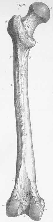
a) Head of the femoral bone (bound to the Acetabulum – an enarthosis).
b) Femoral head fossa for the round ligament.
c) Femoral neck.
d) Greater trochanter (insertion site for mm gluteus medius and minimus, piriformis,
gemelli, obturator and quadratus femoris).
e) Lesser trochanter (insertion site for mm psoas major and internal iliac).
f) Posterior intertrochanteric line (insertion for m quadratus femoris).
g) Upper limb of the linea aspera.
h) Linea aspera (insertion for pecineus, gluteus maximus, femoral adductors,
vasti and adductor brevis).
i) Lower limb of the linea aspera.
k) Femoral body.
l) Popliteal fossa.
m) Lateral condyle.
n) Medial condyle.
o) Intercondylar fossa.