For Providers -- Atlas of Human Anatomy
Translated by: Ronald A. Bergman, PhD and Adel K. Afifi, MD, MS
Peer
Review Status: Internally Peer Reviewed
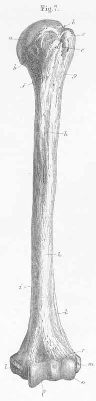
a) Humeral head.
b) Humeral neck.
c) Greater tubercle (insertion for mm supra- and infraspinatus and teres minor).
d) Lesser tubercle (insertion for m subcapsularis).
e) Intertubercular sulcus (passage for tendon for long head of biceps).
f) Crest of the lesser tubercle (insertion mm teres major, lattisimus dorsi,
and coracobrachialis).
g) Crest of the greater tubercle (insertion for mm deltoideus and pectoralis
major.
h) Body, diaphysis of humerus (origin for m brachialis).
i) Medial margin.
k) Insertion of muscle brachioradialis.
l) Medial epicondyle (origin of mm flexores and m palmaris longus).
m) Lateral epicondyle.
n) Humeral elbow joint.
o) Trochlea of humerus.
p) Trochlear groove.
q) Capitulum.
r) Radial fossa.