For Providers -- Atlas of Human Anatomy
Translated by: Ronald A. Bergman, PhD and Adel K. Afifi, MD, MS
Peer
Review Status: Internally Peer Reviewed
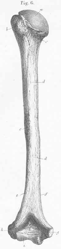
a) Head of the humerus.
b) Greater tubercle of humerus.
c) Neck of the humerus (insertion of capsular ligament).
d) Body of the humerus, diaphysis.
e) Groove for radial nerve.
f) Margin of medial angle (origin for m. triceps, medial head).
g) Medial condyle (flexor) (with groove for ulnar nerve and origin for mm pronator
teres, flexor carpi radialis and ulnaris, palmaris longus, flexor digitorum
superficialis and ligament laterale internum).
h) Lateral condyle (extensor) (origin for mm triceps, lateral head, supinator
longus and brevis, extensor carpi radialis and brevis, extensors digitorum and
for ligament laterale externum).
i) Trochlea of humerus.
k) Olecranon fossa.