For Providers -- Atlas of Human Anatomy
Translated by: Ronald A. Bergman, PhD and Adel K. Afifi, MD,
MS
Peer Review Status: Internally Peer Reviewed
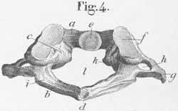
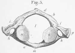
a) Anterior arch of the atlas (attachment site for rectus capitis
anterior minor, anterior atlanto-occipital ligament).
b) Posterior arch.
c) Lateral mass.
d) Posterior tubercle of the atlas (origin of rectus capitis
posterior minor).
e) Fovea dentis atlantis.
f) Condyloid fossa of the superior oblique process (formed by the
condyle of the occipital bone; a hinge joint).
g) Transverse atlantic process (attachment site for mm rectus capitis
lateralis, superior and inferior capitis oblique).
h) Vertebral foramen (transverse foramen) (vertebral canal for the
vertebral artery.
i) Groove for the vertebral artery.
k) Lateral tubercle for ligamentum transversum. (lig. Cruciforme
atlantis).
l) Vertebral foramen or canal.
m) Anterior atlantic tubercle (attachment for m. longus colli, and
anterior longitudinal ligament).
n) Superficial articular site for the axis.