For Providers -- Atlas of Human Anatomy
Translated by: Ronald A. Bergman, PhD and Adel K. Afifi, MD, MS
Peer
Review Status: Internally Peer Reviewed
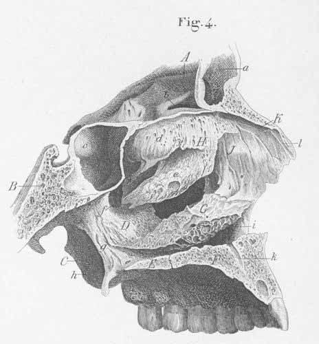
A) Frontal bone, frontal part.
B) Sphenoid bone, body.
C) Pterygoid process of the sphenoid bone.
D) Palatine bone, perpendicular part.
E) Palatine bone, horizontal part.
F) Maxillary bone, palatine process.
G) Inferior concha, inferior turbinate bone.
H) Nasal lamina, ethmoid labyrinth.
I) Frontal process (nasalis of the maxillary bone).
K) Nasal bone.
a) Frontal sinus.
b) Ethmoidal sinus.
c) Sphenoidal sinus.
d) Superior concha, highest turbinate bone.
e) Middle concha, middle turbinate bone.
f) Pterygopalatine foramen, in the perpendicular part of the palatine bone.
g) Pterygoid process, medial wing.
h) Pterygoid process, lateral wing.
i) Lacrymal canal, exit.
k) Incisive canal (for the passage of the nasopalatine artey, vein, and nerve).
l) Sulcus for the external nasal branch of the anterior ethmoid nerve.