For Providers -- Atlas of Human Anatomy
Translated by: Ronald A. Bergman, PhD and Adel K. Afifi, MD, MS
Peer
Review Status: Internally Peer Reviewed
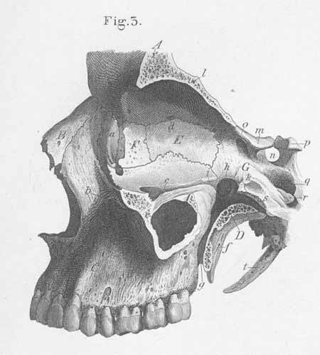
A. Frontal bone.
B. Nasal bone
C. Maxillary bone.
D. Palatine bone (perpendicular part; pterygopalatine fossa).
E. Ethmoidal bone (lamina papyracea).
F. Lacrymal bone.
G. Sphenoid bone.
a) Fossa for lacrymal sac.
b) Infraorbital foramen.
c) Ethmoidal foramen.
e) Maxillary sinus (antrum of Highmore).
f) Pterygoid process.
g) Pterygopalatine canal.
h) Pterygopalatine foramen.
i) Orbital process, palatine bone.
k) Sphenoid process, palatine bone.
l) Orbital part of the frontal bone.
m) Anterior clinoid process.
n) Sella turcica.
o) Optic foramen.
p) Posterior clinoid process.
q) Carotid canal.
r) Lingula.
s) Pterygoid canal (Vidian canal).
t) Styloid process.