ความคิดเห็นทั้งหมด : 8

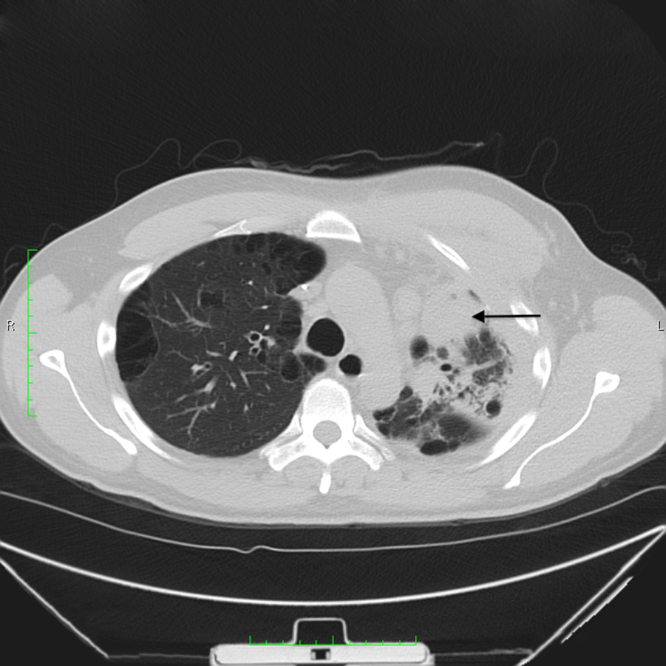
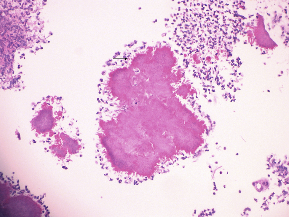
ชายอายุ 45 ปีมีเต้านมข้างซ้ายบวมแข็งและเจ็บ+มีไข้หนาวสั่น มา 1 เดือน


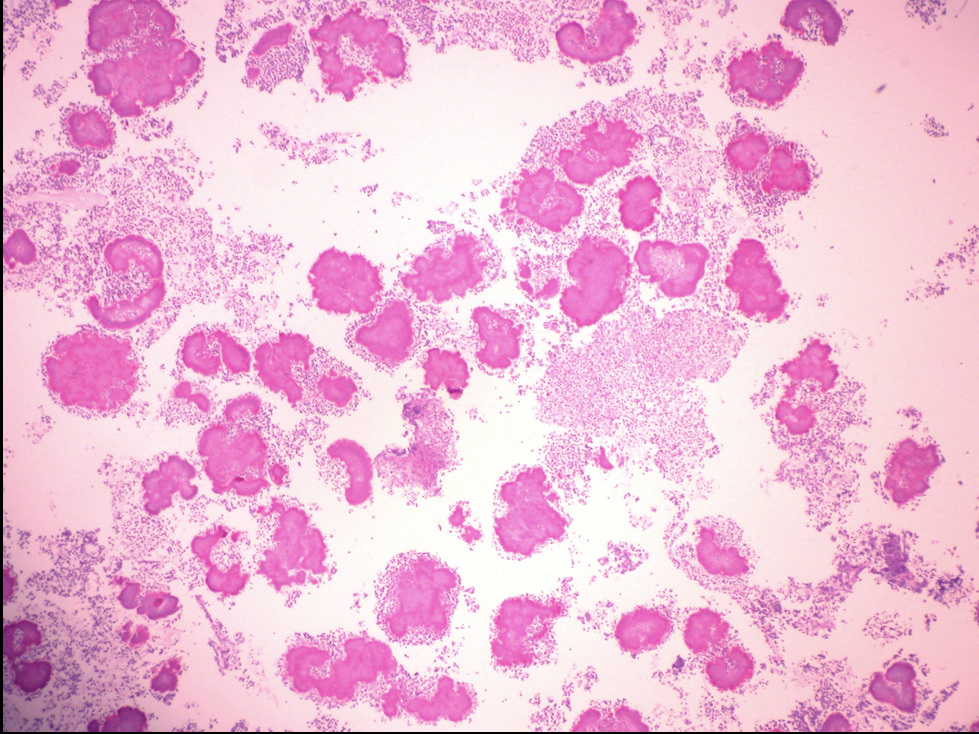
   ชายอายุ 45 ปีมีเต้านมข้างซ้ายบวมแข็งและเจ็บร่วมกับมีไข้หนาวสั่น มา 1 เดือน.
PE: afebrile, Oxygen sat 99% on room air. Left anterior chest wall : a hyperpigmented, firm, and indurated mass inferior to the nipple, extending to the left lateral wall. The mass was exquisitely tender to touch.
Labs: WBC 20,200/cu mm. CT chest (รูป 1) delineated the mass as involving the left breast and pectoralis muscle, with extension into the left chest resulting in pleural thickening and significant airspace density in the left upper lobe. Mediastinal lymphadenopathy was also noted.
ผู้ป่วยได้รับการทำ core biopsy และ aspiration of the mass. เจาะได้ purulent fluid หลายซีซี.


Posted by : cpantip , E-mail : (chpantip@gmail.com) ,
Date : 2018-05-03 , Time : 14:36:00 , From IP : 172.29.3.190


ความคิดเห็นที่ : 1


   รูป 2 CT chest ของผู้ป่วย
รูป 1 คือ histologic section ของ biopsy.
1. การวินิจฉัยคืออะไร
2. จะ manage อย่างไร


Posted by : cpantip , Date : 2018-05-03 , Time : 14:38:52 , From IP : 172.29.3.190

ความคิดเห็นที่ : 2


    รูป 1 แสดงถึง multiple sulfur granules. Sulfur granules นี้ประกอบด้วย colonies ของเชื้อหลายๆ colonies. รูป 3 แสดง higher magnification ของ sulfur granule อันหนึ่ง ซึ่ง เห็น eosinophilic border ที่มีชื่อเรียกว่า Splendore-Hoeppli phenomenon (ลูกศร)ได้อย่างชัดเจน.


Posted by : cpantip , Date : 2018-05-03 , Time : 14:41:09 , From IP : 172.29.3.190

ความคิดเห็นที่ : 3


   การวินิจฉัย: Actinomycosis caused by Actinomyces israelii จาก pathology
แต่ผลเพาะเชื้อของน้ำที่เจาะได้ขึ้น Actinobacillus actinomycetemcomitans จะ manage อย่างไรต่อไป


Posted by : cpantip , Date : 2018-05-03 , Time : 14:41:59 , From IP : 172.29.3.190

ความคิดเห็นที่ : 4


    ตั้งแต่แรก แพทย์คิดว่าผู้ป่วยเป็น CA breast แต่มาให้การวินิจฉัย actinomycosis จากผลของ pathology แพทย์จึงเริ่มให้การรักษาด้วย PGS (5 million units IV ทุก 6 ชั่วโมง).
อย่างไรก็ตาม เนื่องจาก breast fluid culture ขึ้นเชื้อ Actinobacillus actinomycetemcomitans, original diagnosis จึงยังเป็นปัญหา.


Posted by : cpantip , Date : 2018-05-03 , Time : 14:42:38 , From IP : 172.29.3.190

ความคิดเห็นที่ : 5


    Actinobacillus actinomycetemcomitans เป็นหนึ่งในเชื้อหลายตัวที่สามารถเพาะเชื้อขึ้นได้ร่วมกับ Actinomyces species โดยพบ Actinobacillus actinomycetemcomitans อยู่ใน 30% ของรอยโรคของ actinomycosis.


Posted by : cpantip , Date : 2018-05-03 , Time : 14:43:21 , From IP : 172.29.3.190

ความคิดเห็นที่ : 6


    เนื่องจากพบ sulfur granules จำนวนมากใน pathology และเพราะผู้ป่วยมี classic presentation, Actinomyces species จึงยังคงเป็น most likely primary pathogen. ผู้ป่วยได้รับ penicillin ต่อไปโดยมีอาการดีขึ้นอย่างรวดเร็ว. อีก 7 วันต่อมาanaerobic cultures ขึ้นเชื้อ A. israelii. ผู้ป่วยรายนี้หายโดยได้รับยา penicillin เพื่อรักษาการติดเชื้อ actinomyces อย่างเดียว.


Posted by : cpantip , Date : 2018-05-03 , Time : 14:44:39 , From IP : 172.29.3.190

ความคิดเห็นที่ : 7


   Actinomyces species เป็น gram-positive, non-spore-forming และ predominantly anaerobic bacteria. เชื้อนี้เป็น commensal ของ human oropharynx, gastrointestinal tract และ female genitalia. การวินิจฉัย actinomycosis มักจะล่าช้าจนกระทั่งได้ทำ biopsy เพื่อการประเมิน malignancy. ผู้ป่วยรายนี้มี periodontal disease. Thoracic disease ของเขาน่าจะเกิดจาก aspiration oropharyngeal secretion มากที่สุด.


Posted by : cpantip , Date : 2018-05-03 , Time : 14:45:14 , From IP : 172.29.3.190

ความคิดเห็นที่ : 8


   Reference: Tang L, et al. Clinical Infectious Diseases 2015;60:438, 486-7.


Posted by : cpantip , Date : 2018-05-03 , Time : 14:45:44 , From IP : 172.29.3.190

สงวนสิทธิ์การแสดงความคิดเห็นสำหรับ สมาชิกเท่านั้น