ความคิดเห็นทั้งหมด : 8

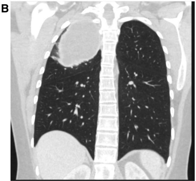
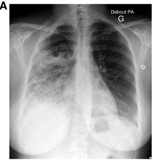
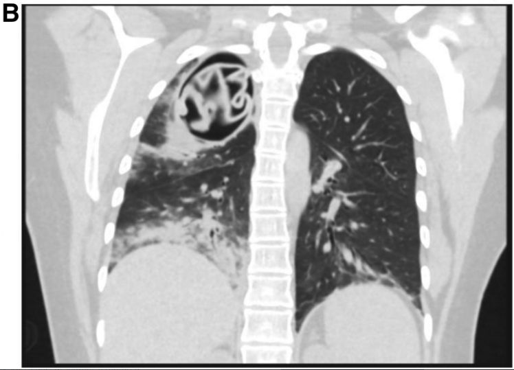
หญิงชาวตุรกีอายุ 27 ปี เจ็บหน้าอกมา 1 เดือน


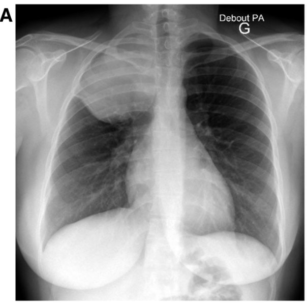
   หญิงชาวตุรกีอายุ 27 ปีมาด้วยอาการเจ็บหน้าอกมา 1 เดือน. At admission, she was afebrile, physical examination revealed reduced vesicular breath sounds of the right upper lobe.
รูป A และ B คือ Chest radiography และ whole body computed tomography scan
1. การวินิจฉัยคืออะไร
2. จะ manage อย่างไร


Posted by : cpantip , E-mail : (chpantip@gmail.com) ,
Date : 2017-11-23 , Time : 11:39:39 , From IP : 172.29.3.70


ความคิดเห็นที่ : 1


   รูป A และ B คือ Chest radiography และ whole body computed tomography scan

Posted by : cpantip , Date : 2017-11-23 , Time : 11:40:29 , From IP : 172.29.3.70

ความคิดเห็นที่ : 2


    Chest radiography และ whole body computed tomography scan showed a thoracic cyst-like mass with smooth borders, no calcification, and ruled out intraabdominal lesion (Figure 1A and B).


Posted by : cpantip , Date : 2017-11-23 , Time : 11:41:17 , From IP : 172.29.3.70

ความคิดเห็นที่ : 3


    แพทย์นึกถึงEchinococcus granulosus infection เนื่องจาก positive Western Blot against p7 and p26-kD bands. แพทย์ได้วางแผนทำ lobectomy อีก 1 เดือน และผู้ป่วยได้กลับบ้าน.

Posted by : cpantip , Date : 2017-11-23 , Time : 11:41:46 , From IP : 172.29.3.70

ความคิดเห็นที่ : 4


    3 สัปดาห์ต่อมา ผู้ป่วยมีอาการเจ็บหน้าอกขึ้นมาทันที ร่วมกับมีไข้และไอ. ตรวจ CBC พบ eosinophilia (3,270 mm3) และ elevated C-reactive protein level (226 mg per litter).
CXR ดังในรูป


Posted by : cpantip , Date : 2017-11-23 , Time : 11:42:30 , From IP : 172.29.3.70

ความคิดเห็นที่ : 5


   Chest radiography (Panel A) and computed tomography scan (Panel B) showing a ruptured hydatid cyst with underneath interstitial pneumonia and apparition of a pathognomonic germinative membrane.

Posted by : cpantip , Date : 2017-11-23 , Time : 11:43:13 , From IP : 172.29.3.70

ความคิดเห็นที่ : 6


    ผู้ป่วยได้รับการผ่าตัด lobectomy ที่ 1 สัปดาห์หลังจากได้รับการรักษาด้วยยา albendazole.
การผ่าตัดพบ an empty cyst ซึ่งได้รับการทำ totally removed และ a bronchial fistula ซึ่งถูกเย็บปิด. Pathology analysis ยืนยันการมี Echinococcus granulosus protoscolex และ rostellar hooks. ผู้ป่วยได้รับการรักษาด้วย albendazole 15 mg/kg per day ต่อเนื่องไปนาน 6 เดือน.
จากการติดตามที่นาน 3 ปี ไม่มี relapse และ chest radiography ปกติ.


Posted by : cpantip , Date : 2017-11-23 , Time : 11:43:45 , From IP : 172.29.3.70

ความคิดเห็นที่ : 7


    Echinococcosis เป็น cestodiasis ที่เกิดจากการติดเชื้อด้วย larval stage ของ Echinococcus. มีอยู่4 species ของ Echinococcus ที่พบในคน คือ: E. granulosus ที่ทำให้เกิด cystic echinococcosis (CE) และ Echinococcus multilocularis ที่ทำให้เกิด alveolar echinococcosis, ทั้ง 2 species พบได้บ่อยที่สุด. อีก 2 species คือ Echinococcus vogeli และ Echinococcus oligarthrus ทำให้เกิด polycystic echinococcosis ซึ่งพบน้อยมากในคน. E. granulosus เป็นสาเหตุที่พบบ่อยที่สุดของโรคที่เป็น unilocular cystic lesions ซึ่งมักจะเกิดขึ้นที่ตับและปอด แต่ก็เกิดขึ้นได้ที่อวัยวะอื่นๆ. แม้ว่าการติดเชื้อนี้ ส่วนใหญ่ผู้ป่วยไม่มีอาการใดๆ โดย CE ทำให้เกิด cysts ที่โตขึ้นช้าๆ ใช้เวลานานหลายปีโดยผู้ป่วยไม่ได้สังเกต จนกระทั่งเกิด acute complications เช่น cyst rupture ทำให้เกิดไอ เจ็บหน้าอก ไอเป็นเลือดได้. การผ่าตัดยังเป็นการรักษาหลักเพื่อตัดเอา cyst ออกไป และเย็บปิด bronchial fistula ถ้าจำเป็น ร่วมกับการให้ยาขับพยาธิเป็นระยะยาวนาน.

Posted by : cpantip , Date : 2017-11-23 , Time : 11:44:15 , From IP : 172.29.3.70

ความคิดเห็นที่ : 8


    Reference: Argemi X, et al. Pulmonary cystic echinococcosis. Am J Trop Med Hyg 2017;97 (3):641 –2.

Posted by : cpantip , Date : 2017-11-23 , Time : 11:45:02 , From IP : 172.29.3.70

สงวนสิทธิ์การแสดงความคิดเห็นสำหรับ สมาชิกเท่านั้น