ความคิดเห็นทั้งหมด : 12

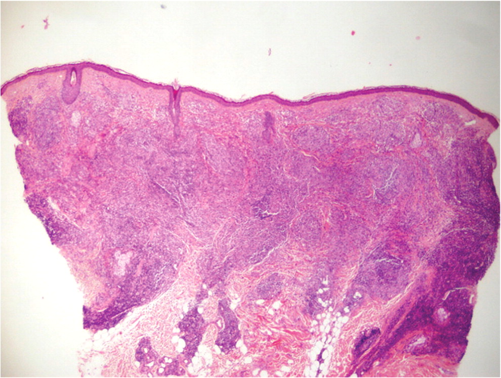
หญิงอายุ 45 ปี มีรอยโรคที่แก้มข้างซ้ายมานาน 2 ปี.


   หญิงอายุ 45 ปีมาโรงพยาบาลเนื่องจากมีรอยโรคที่แก้มข้างซ้ายมานาน 2 ปี. ผู้ป่วยไม่มีโรคประจำตัวใดๆ.
ตรวจร่างกายพบความผิดปกติดังในรูป 1.
1. ท่านต้องการตรวจร่างกายอะไรเพิ่มเติมอีก
2. การวินิจฉัยน่าจะเป็นโรคใด
3. จะ manage อย่างไร


Posted by : cpantip , E-mail : (chpantip@gmail.com) ,
Date : 2016-06-27 , Time : 11:17:56 , From IP : 172.29.3.247


ความคิดเห็นที่ : 1


   Korapin Settasuporn ตอบมา
1.ตรวจการชาของใบหน้า , ตรวจว่ามีเส้นประสาทโตหรือไม่ค่ะ
ตรวจว่ามีผื่นลักษณะนี้ที่เดียว หรือบริเวณอื่นด้วยหรือไม่ค่ะ
2.โรคเรื้อนค่ะ
3.ส่ง biopsy ตรงlesion ไปตรวจ acid fast bacili ค่ะ
ให้การรักษาตามระดับของโรคเชื้อน้อย/เชื้อมาก
และ เเจ้งสำนักระบาดวิทยาค่ะ


Posted by : cpantip , Date : 2016-06-27 , Time : 11:19:01 , From IP : 172.29.3.247

ความคิดเห็นที่ : 2


   PE: a gross infiltrated erythematous and scaly plaque with poorly defined edges that measured 18 cm in diameter. Her left earlobe was thickened and anesthetic. Satellite erythematous papules were observed on her neck. There was no peripheral nerve enlargement.
แพทย์ได้ทำ biopsy ที่ขอบของรอยโรคอันใหญ่แล้วส่งไปทำ routine hematoxylin-eosin, Ziehl-Nielsen, Fite-Faraco และ periodic acid-Schiff staining (รูป 2 และ 3) และส่งเพาะเชื้อ bacteria, mycobacteria และ fungus


Posted by : cpantip , Date : 2016-06-27 , Time : 11:20:41 , From IP : 172.29.3.247

ความคิดเห็นที่ : 3


   Fite-Faraco stain

Posted by : cpantip , Date : 2016-06-27 , Time : 11:22:27 , From IP : 172.29.3.247

ความคิดเห็นที่ : 4


   การวินิจฉัย: multibacillary borderline lepromatous disease.

Posted by : cpantip , Date : 2016-06-27 , Time : 11:23:14 , From IP : 172.29.3.247

ความคิดเห็นที่ : 5


   Biopsy specimens จาก erythematous, scaly plaque ที่แก้มของผู้ป่วยพบว่าเป็น diffused epithelioid granulomas with a peripheral rim of lymphocytes distributed throughout the dermis and subcutis and elongated along the course of the vessels (รูป 2). There were abundant acid-fast bacilli singly or in clumps inside the bubbly histiocytes visible on Fite-Faraco staining (รูป 3). No nerves were identified by hematoxylin-eosin and S100 protein staining.


Posted by : cpantip , Date : 2016-06-27 , Time : 11:24:06 , From IP : 172.29.3.247

ความคิดเห็นที่ : 6


   Hematoxylin and eosin stain showing multiple elongated granulomas distributed throughout the dermis and subcutis (arrows) (original magnification, ×40).

Posted by : cpantip , Date : 2016-06-27 , Time : 11:24:34 , From IP : 172.29.3.247

ความคิดเห็นที่ : 7


   Fite-Faraco stain showing abundant acid-fast bacilli singly or in clumps inside the bubbly histiocytes (arrows) (original magnification, ×1000).

Posted by : cpantip , Date : 2016-06-27 , Time : 11:25:08 , From IP : 172.29.3.247

ความคิดเห็นที่ : 8


   ผู้ป่วยได้รับการรักษาด้วย clofacimine (50 mg per day), rifampicin (600 mg per month), and dapsone (100 mg per day) หลังจากรักษาไปได้นาน 1 ปี รอยโรคดีขึ้นอย่างมาก. อย่างไรก็ตาม ผู้ป่วยต้องได้รับยาต่อไปอีก 1 ปี.

Posted by : cpantip , Date : 2016-06-27 , Time : 11:25:47 , From IP : 172.29.3.247

ความคิดเห็นที่ : 9


   Leprosy เป็น chronic granulomatous infection ซึ่งเป็นที่ผิวหนังและ peripheral nerves เป็นหลัก. เชื้อก่อโรคนี้คือ obligate intracellular organism Mycobacterium leprae. Leprosy มีรูปแบบที่หลากหลายซึ่งสัมพันธ์กับ bacterial index และ cell-mediated immunity ซึ่งทำให้ผู้ป่วยมี clinicopathological manifestations แตกต่างกัน (เป็น polar tuberculoid หรือ lepromatous, borderline tuberculoid หรือlepromatous, mid-borderline และ pure neuritic forms).


Posted by : cpantip , Date : 2016-06-27 , Time : 11:26:28 , From IP : 172.29.3.247

ความคิดเห็นที่ : 10


   Borderline leprosy มีรอยโรคของผิวหนังที่อยู่ระหว่าง 2 polar forms. Morphology ของรอยโรคอาจจะเป็น macular, papulonodular, plaque-like, annular หรือมี geographic appearance. Infiltration ที่มีหรือไม่มี nodulation เหนือ pinna, เด่นที่ helixเป็นลักษณะเฉพาะของ lepromatous leprosy. External ear involvement อาจมีเพียงเล็กน้อย, เป็น diffuse infiltration หรือ discrete nodules, หรือ extensive. อาจพบแผลที่เหมือนมีรอยตอด หรือหนูแทะ (“nibbled” or “rat-bitten” defect). หูอาจมีลักษณะเป็น megalobule จาก earlobe ยืดยาว เหี่ยวย่นและห้อยลงมา. อาจพบ auricular chondritis :ซึ่งเป็นส่วนหนึงของ reactional state.


Posted by : cpantip , Date : 2016-06-27 , Time : 11:27:07 , From IP : 172.29.3.247

ความคิดเห็นที่ : 11


   การ form เป็น small granulomas เป็นลักษณะเฉพาะของ borderline leprosy, และ granulomas กระจายออกไปเมื่อ condition เปลี่ยนจาก borderline tuberculoid ไปเป็น borderline lepromatous disease. มี acid-fast bacilli มากใน lepromatous lesions, แต่ไม่พบ acid-fast bacilli ใน tuberculoid lesions. การตรวจพบ acid-fast bacilli มีประโยชน์ในการวินิจฉัย, เพราะ borderline states เป็น immunologically unstable, and laboratory test results สามารถถูกทำให้ซับซ้อนได้จาก reactions (เช่น type 1 lepra reaction, type 2 lepra reaction และ Lucio reaction).

Posted by : cpantip , Date : 2016-06-27 , Time : 11:27:20 , From IP : 172.29.3.247

ความคิดเห็นที่ : 12


   Reference: Gomez-Moyano E, et al. Large infiltrated erythematous and scaly plaque on the cheek and thickened earlobe. Clin Infect Dis 2010; 50 (7): 1015-6, 1068-9.

Posted by : cpantip , Date : 2016-06-27 , Time : 11:27:46 , From IP : 172.29.3.247

สงวนสิทธิ์การแสดงความคิดเห็นสำหรับ สมาชิกเท่านั้น