ความคิดเห็นทั้งหมด : 7

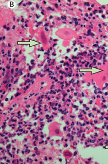
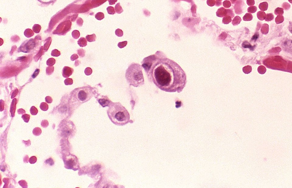
ชายอายุ 61 ปี มี esophageal lesion


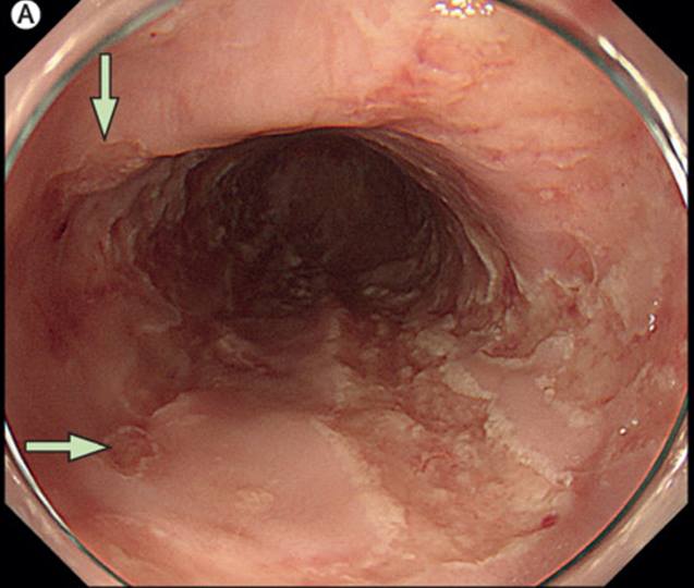
   ชายอายุ 61 ปี ซึ่งมีประวัติ cord-blood transplantation สำหรับ acute myeloid leukemia เมื่อ 15 ปีก่อน และ chronic graft-versus-host disease ของผิวหนังซึ่งได้รับการรักษาด้วย maintenance dose คือ prednisone 5 mg/วัน ได้เข้ารับการรักษาในโรงพยาบาลเนื่องจากมี upper GI bleeding จาก duodenal ulcer. ผู้ป่วยได้รับการรักษาด้วย combination ของ endoscopic clipping proton-pump inhibitor ซึ่งได้ผลดี. 7 วันหลัง GI bleeding แพทย์ได้ทำ EGD scopy ซ้ำ พบว่ามี multiple, newly developed, shallow ulcers in the esophagus. Some of the lesions were circumscribed and discrete (รูป A).


Posted by : cpantip , E-mail : (chpantip@gmail.com) ,
Date : 2016-05-31 , Time : 14:44:01 , From IP : 172.29.3.57


ความคิดเห็นที่ : 1


   รูป B คือ histopathology of the esophagus.

การวินิจฉัย คืออะไร


Posted by : cpantip , Date : 2016-05-31 , Time : 14:44:33 , From IP : 172.29.3.57

ความคิดเห็นที่ : 2


   ตอบ HSV esophagitis

รูป (A) Endoscopic findings of esophagitis due to herpes simplex virus. Arrows indicate volcano-like ulcers in the esophagus
รูป (B) Histopathological findings of esophagitis: arrows indicate multinucleated epithelial giant cells with eosinophilic intracellular inclusion bodies, consistent with Cowdry type-A inclusion body.

ใน biopsy samples ไม่พบ malignancy หรือ graft-versus-host disease. Immunohistochemical staining of the specimens ได้ผลบวกสำหรับ herpes simplex virus, แต่ได้ผลลบสำหรับ cytomegalovirus.


Posted by : cpantip , Date : 2016-05-31 , Time : 14:45:52 , From IP : 172.29.3.57

ความคิดเห็นที่ : 3


   ผู้ป่วยได้รับการรักษาด้วย intravenous acyclovir นาน 10 วันแล้วแพทย์ได้ทำ EGD scopy ซ้ำ ซึ่งพบว่าหายดี.
Herpes simplex virus เป็นหนึ่งในเชื้อที่ทำให้เกิด infective esophagitis ได้บ่อยที่สุด.
Herpes simplex esophagitis พบได้บ่อยในผู้ป่วยที่มี immunosuppressive status ซึ่งรวมถึง หลัง bone marrow transplantation แต่ก็พบได้ในคนที่ immunocompetent. Esophagitis ในผู้ป่วยเหล่านี้ก็อาจเกิดจากเชื้ออื่นๆ เช่น cytomegalovirus หรือ Candidia spp.


Posted by : cpantip , Date : 2016-05-31 , Time : 14:46:56 , From IP : 172.29.3.57

ความคิดเห็นที่ : 4


   การวินิจฉัย herpes simplex esophagitis อาศัยการยืนยันจาก combination ของ gross EGD scopy findings, a histopathological examination และ immunohistochemical stain ของ biopsied specimen.

Esophageal herpes simplex lesions ที่พบจาก EGD scopy ที่ typical เรียกว่า volcano ulcers มีลักษณะเป็น discrete, diffusely scattered shallow ulcers. The Cowdry type-A inclusion body เป็น classic histopathological finding ใน biopsy specimens ของ herpes simplex esopohagitis ดังที่พบในผู้ป่วยรายนี้ และพบได้ถึง 70% ของ biopsy specimens.


Posted by : cpantip , Date : 2016-05-31 , Time : 14:48:05 , From IP : 172.29.3.57

ความคิดเห็นที่ : 5


    Reference: Takeuchi Y, et al. Volcano-like shallow oesophageal ulcers in a patient with a history of cord-blood transplantation. Lancet Infect Dis 2016;16(3):384.


Posted by : cpantip , Date : 2016-05-31 , Time : 14:50:23 , From IP : 172.29.3.57

ความคิดเห็นที่ : 6


   CMV
Description of CMV:
Characterized by intra-nuclear inclusion with enlargement of both cell & nucleus. Cytoplasmic inclusions are also seen in the infected cells.
Nuclear inclusion: Single, measuring up to 20 µm in diameter, while the cytoplasmic inclusions are much smaller, measuring 1 to 3 µm.
It is usually basophilic but may be eosinophilic, round to oval.


Posted by : cpantip , Date : 2016-05-31 , Time : 14:51:27 , From IP : 172.29.3.57

ความคิดเห็นที่ : 7


   CMV

Posted by : cpantip , Date : 2016-05-31 , Time : 14:51:49 , From IP : 172.29.3.57

สงวนสิทธิ์การแสดงความคิดเห็นสำหรับ สมาชิกเท่านั้น