ความคิดเห็นทั้งหมด : 4

ชายอายุ 72 ปี เป็น type 2 DM ปวดท้อง และมีความผิดปกติที่กระดูก


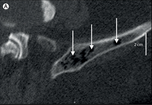
   ชายอายุ 72ปีซึ่งเป็น type 2 DM มาพบแพทย์เนื่องจากมีอาการปวดท้องหลังจากหกล้มลำตัวด้านขวากระแทกพื้น. ผู้ป่วยได้รับ anticoagulants อยู่เนื่องจากมี atrial fibrillation. CT abdomen พบ hepatic haematoma. ตรวจพบ ค่า C-reactive protein และ procalcitonin concentrations สูง ซึ่งแสดงว่าน่าจะมี superinfection. ผู้ป่วยได้รับการทำ diagnostic aspiration และได้รับการรักษาด้วย IV ceftriaxone. วันรุ่งขึ้น อาการของผู้ป่วยทรุดลง แพทย์ได้เปลี่ยนยาเป็น meropenem.
Follow-up CT scan พบความผิดปกติที่ pelvis และ lumbar vertebra ดังในรูป A.

1. ความผิดปกติที่กระดูกนี้คืออะไร
2. จะ manage อย่างไร


Posted by : cpantip , E-mail : (chpantip@gmail.com) ,
Date : 2015-04-15 , Time : 16:50:50 , From IP : 172.29.3.187


ความคิดเห็นที่ : 1


   พบ gas ใน bone ค่ะ จากอาการ+อาการแสดงของผู้ป่วย ความผิดปกตินี้น่าจะเกิดจากการติดเชื้อมากที่สุด การวินิจฉัยคือ emphysematous osteomyelitis เชื้อก่อโรคที่ทำพยาธิสภาพแบบนี้ต้องเป็นเชื้อที่สร้าง gas ได้ คือ เชื้อกลุ่ม anaerobe และ enterobacteriaceae การเกิด emphysematous osteomyelitis ในผู้ป่วยรายนี้ น่าจะมาทาง hematogenous
การรักษา 1. antibiotic ให้ meropenem ก็เหมาะสมดีอยู่แล้วเพราะครอบคลุม เชื้อกลุ่ม anaerobe และ enterobacteriaceae
2. หา source of infection เพื่อดูว่าจะ remove source of infection ได้อย่างไรบ้าง


Posted by : daisy1 , Date : 2015-04-17 , Time : 15:40:52 , From IP : 172.29.3.187

ความคิดเห็นที่ : 2


   Follow-up CT scan พบ intraosseous gas ใน pelvis และ lumbar vertebra (figure A).

CT-guided bone biopsy ยืนยันว่ามี gas อยู่ในกระดูก (figure B), เข้าได้กับ emphysematous osteomyelitis, และแพทยืได้เพิ่มยา clindamycin เข้าไปอีก.

ผล bone biopsy culture ขึ้น non-resistant Escherichia coli. Microbiological culture ของ liver sample, urine และ catheter tips และ arterial และ venous blood cultures ไม่ขึ้นเชื้อใดๆ.

ทั้งๆ ที่ให้ IV antibiotic ขนาดสูง ผู้ป่วยเสียชีวิตจาก multiorgan failure จาก fulminant sepsis โดยไม่พบ source ของ infection ที่ใด.


Posted by : cpantip , Date : 2015-04-22 , Time : 08:16:40 , From IP : 172.29.3.187

ความคิดเห็นที่ : 3


   Figure.
Evidence of intraosseous gas on CT and histopathology
(A) Unenhanced CT image using CT bone window settings; arrows point to the sites of intraosseous gas. (B) Microphotograph of histopathology section (periodic acid–Schiff stain) obtained during bone biopsy; asterisks show sites of intraosseous gas.


Posted by : cpantip , Date : 2015-04-22 , Time : 08:17:38 , From IP : 172.29.3.187

ความคิดเห็นที่ : 4


   การพบว่ามี intraosseous gas มี differential diagnosis คือ vacuum phenomena, subchondral cysts ใน degenerative joint disease, penetrating (รวมถึง iatrogenic) injury, ischemic necrosis และ osteomyelitis ที่เกิดจาก gas-forming organisms.

Pattern ของ distribution, detailed appearance, clinical history, และ findings ในการตรวจในการนำมาซึ่งการวินิจฉัย. ใน systemic illness ที่มี septicemia สามารถใช้ CT scanning เพื่อ localise ตำแหน่งของการติดเชื้อ และเพื่อ staging microbial invasion และ host response. เชื้อที่สร้างแก้สเมื่อ colonising bone คือ Pseudomonas spp, Streptococcus spp, Enterococcus spp, E coli, Salmonella spp, Staphylococcus aureus และ fungi.

ใน emphysematous osteomyelitis, gas ปรากฎขึ้นจากการรวมตัวของฟองอากาศเล็กๆ ที่อยู่ภายใน medullary cavity โดยมี narrow zone ของ transition และไม่มี sclerotic rim.

การมี intraosseous gas ในผู้ป่วยที่มี systemic infection ผุ้ป่วยอาจไม่แสดงอาการทางคลินิกใดๆ ให้นึกถึงภาวะนี้ แต่ควรมองหาความผิดปกตินี้ไว้ด้วยเวลาที่ดู CT scans ของผู้ป่วย.

Reference: Larsen J, et al. Emphysematous osteomyelitis.Lancet ID 2015;15:485.


Posted by : cpantip , Date : 2015-04-22 , Time : 08:24:58 , From IP : 172.29.3.187

สงวนสิทธิ์การแสดงความคิดเห็นสำหรับ สมาชิกเท่านั้น