ความคิดเห็นทั้งหมด : 3

พ่อค้าหมูอายุ 25 ปี ตาซ้ายมองไม่เห็นมา 6 เดือน


   พ่อค้าหมูซึ่งมีสุขภาพดีอายุ 25 ปี มาพบแพทย์เนื่องจากตาซ้ายมองไม่เห็นมา 6 เดือน. ผู้ป่วยไม่ปวดตาเลย.
Visual acuity was 20/20 in the right eye and light perception in the left.

A still retinal image : ดังในรูป B

1. การวินิจฉัยคืออะไร
2. จะ manage อย่างไร


Posted by : cpantip , E-mail : (chpantip@gmail.com) ,
Date : 2015-04-02 , Time : 13:36:28 , From IP : 172.29.3.187


ความคิดเห็นที่ : 1


   Diagnosis : Intraretinal cysticercosis
Management : Albendazole + corticosteroid

ขอบคุณครับ ^^


Posted by : Dejacques , Date : 2015-04-03 , Time : 15:30:47 , From IP : 172.29.3.103

ความคิดเห็นที่ : 2


   คุณหมอ Dejacques ให้การวินิจฉัยถูกแล้วค่ะ

เมื่อ 3 เดือนก่อนมาตรวจครั้งนี้ ผู้ป่วยได้รับการรักษาด้วย albendazole นาน 3 วัน.

รูป
(A) B-scan ultrasound image of the left eye showing a retinal detachment with a hyperechoic focus representing the scolex of the parasite (S) surround by a hypoechoic cystic space in the retina (arrow).
(B) Still retinal image showing a large cystic space (arrow) with the scolex visualised as a yellow circumscribed lesion (S).

Video imaging showed the intraretinal motility of the Taenia solium parasite in response to bright light.

CT scan of the brain พบรอยโรคที่เข้าได้กับ neurocysticercosis.


Posted by : cpantip , Date : 2015-04-05 , Time : 11:32:25 , From IP : 172.29.3.187

ความคิดเห็นที่ : 3


   Ocular cysticercosis — พบได้ 1-3 % ของผู้ป่วย cysticercosis ทั้งหมด. ผู้ป่วยที่มี ocular cysticercosis อาจเป็นที่ subretinal space, vitreous humor, anterior chamber, conjunctiva หรือ extraocular muscles. อาการที่พบคือ impaired vision, recurrent eye pain หรือ diplopia.

ในผู้ป่วยที่มี subretinal cysticerci ต้องนึกไว้เสมอว่าผู้ป่วยอาจมี neurocysticercosis (NCC) อยู่ด้วย แต่การที่มี cysticerci ใน anterior chamber ของตา จัดเป็น extraneural cysticercosis. แม้ว่าผู้ป่วยจำนวนมากไม่มีอาการใดๆ แต่การอักเสบที่เกิดขึ้นรอบๆ degenerating cysticerci สามารถทำให้เกิดการมองเห็นเสียไปได้จากการทำให้เกิด chorioretinitis, retinal detachment หรือ vasculitis. ดังนั้น ในผู้ป่วยที่มี NCC ทุกรายก่อนเริ่มการรักษา ต้องทำ ophthalmologic examination ก่อนเพื่อหาว่ามี ocular cysticercosis หรือไม่

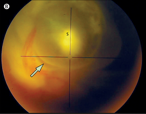
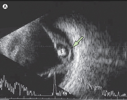
Direct visualization พยาธิโดยตรงด้วย funduscopic examination เป็น pathognomonic สำหรับการวินิจฉัย intraretinal cysticercosis.

การรักษา intraocular cysticercosis ที่ได้รับการรับรองคือ surgical excision. ใน series หนึ่งที่มีผู้ป่วย 45 รายที่มี intraocular cysticercosis, การผ่าตัดสามารถเอา intraocular cysticercosis ออกได้ทุกราย ; ต้องทำ reattachment of the retina ใน 87 % ของ 22 ตา, และ 67% การมองเห็นดีขึ้นจนสามารถช่วยเหลือตัวเองได้.

Reference: Chavala SH, et al. Intraretinal cysticercosis. Lancet 2015;385:799.


Posted by : cpantip , Date : 2015-04-05 , Time : 12:40:56 , From IP : 172.29.3.187

สงวนสิทธิ์การแสดงความคิดเห็นสำหรับ สมาชิกเท่านั้น