ความคิดเห็นทั้งหมด : 12

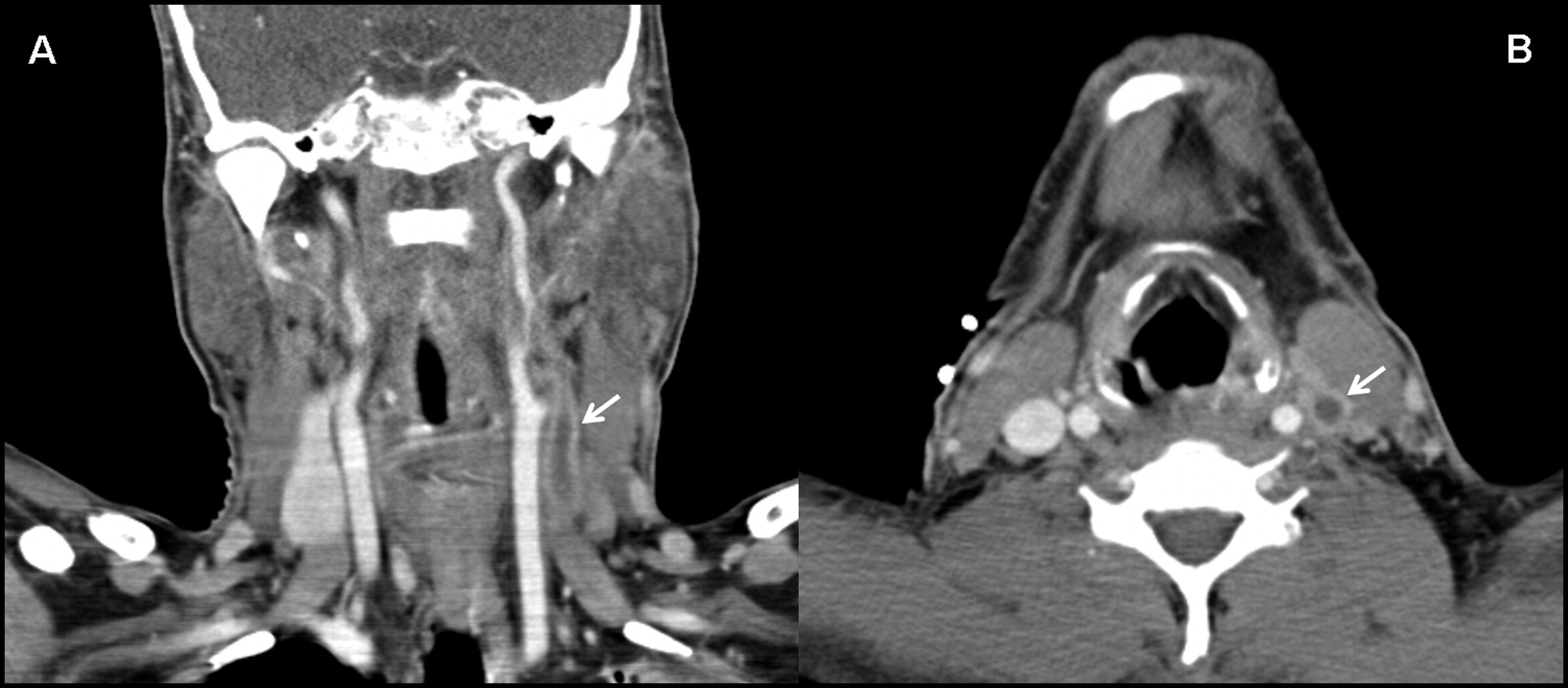
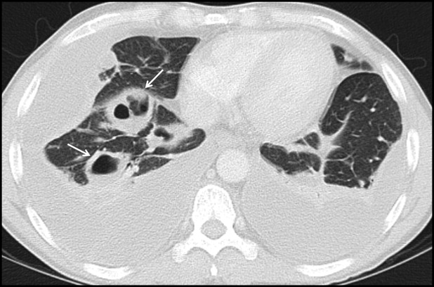
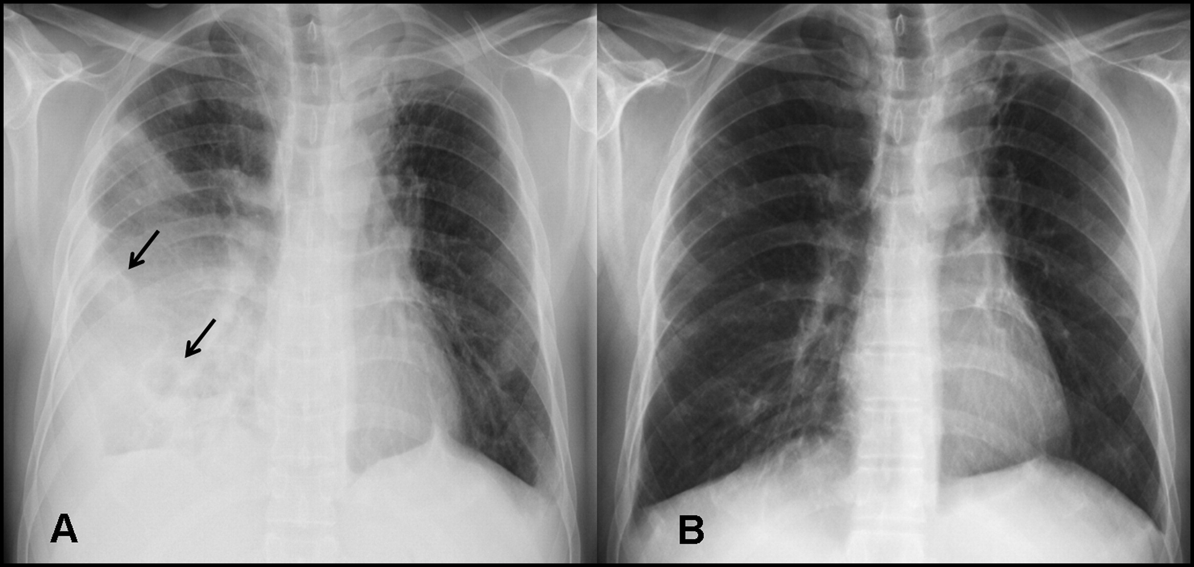
ชายอายุ 39 ปี มีไข้หนาวสั่น เจ็บคอ ไอ เหนื่อยมากขึ้นมา 1 สัปดาห์, BP 80/60, RR 45/min


   ชายอายุ 39 ปี ไม่มีงานทำ ไม่มีบ้าน อาศัยอยู่กับเพื่อน มาที่ห้องฉุกเฉินเพราะมีไข้หนาวสั่นมา 1 สัปดาห์ ร่วมกับเจ็บคอ ไอ เหนื่อยมากขึ้น เสมหะสีน้ำตาล กินอาหารได้น้อยเพราะเบื่ออาหาร
ผู้ป่วยไม่มีโรคประจำตัวใดๆ สูบบุหรี่มานาน (27 pack years) และยังสูบบุหรี่อยู่ ดื่มสุรามาตลอด
PE At presentation, fever, 40.3°C; blood pressure, 80/60 mm Hg; pulse rate, 140 beats per minute; and respiratory rate, 45 breaths per minute.
The submandibular and anterior cervical lymph nodes were enlarged and tender to palpation, and the inspection of the oral cavity revealed an enlargement of the left peritonsillar region with displacement of the uvula to the right. On lung auscultation, predominantly right-sided rales were found; there was no cardiac murmur.

1. ปัญหาแลละการวินิจฉัยคืออะไร
2. จะ manage อย่างไร


Posted by : cpantip , E-mail : (chpantip@gmail.com) ,
Date : 2014-10-23 , Time : 13:00:07 , From IP : 172.29.3.50


ความคิดเห็นที่ : 1


   Problem list
1 septic shock with acute respiratory failure
Source ) deep neck infection ) lt peritonsillar abcess + pneumonia

Mx
On et tube+ mechanical ventilator
Resuscitation
Empiric atb ) ceftriazone + clindamycin cover gram + /- anaerobes ( community setting)
ขอ ct. Head and neck confirm location + extension
Consult ent for adequate drainage ครับ


Posted by : Aekkamon1 , E-mail : (accumulate1985 @gmail.com) ,
Date : 2014-10-23 , Time : 19:14:32 , From IP : cache100.kku.ac.th


ความคิดเห็นที่ : 2


   ขออนุญาตตอบด้วยนะคะ เขียนไว้ตั้งแต่เมื่อคืน เตรียมไว้จะมา post เช้านี้ จะเป็นแนวเดียวกับคุณหมอ Aekkamon1 ค่ะ แต่ละเอียดกว่านิดหน่อย

Pertinent datas
1 Fever with chills, cough with brownish sputum, progressive dyspnea, anorexia
2. Smoking and alcohol drinking
3. T 40.3°C; BP 80/60 mm Hg; P 140/min; R 45 /min = shock, septic shock is the most likely.
4. Left peritonsillar abscess with submandibular and anterior cervical lymphadenitis
5. Lungs: Right-sided rales with marked tachypnea with Hx of fever, cough: pneumonia?

ขณะนี้ผู้ป่วยมี septic shock และ หายใจเร็วมาก อาจมี acute respiratory failure ด้วย
Source of infection คือ left peritoneillar abscess และ pneumonia ซึ่งถ้าคิดเป็นเรื่องเดียวกัน คิดว่าผู้ป่วยมี Lemierre syndrome กล่าวคือ ผู้ป่วยรายนี้เริ่มจาก ไข้+หนาวสั่น+เจ็บคอ ซึ่ง progress ไปเป็น Lt peritonsillar abscess ร่วมกับมี pneumonia ซึ่งน่าจะเกิดจากการติดเชื้อที่ tonsil กระจายไปที่คอทำให้เกิด internal jugular thrombophlebitis แล้วมี septic emboli ไปที่ปอด
เชื้อก่อโรค นึกถึง F. necrophorum มากที่สุดค่ะ

Management
1. วัด O2 sat และให้ oxygen high flow. Evaluate ว่าต้อง endotracheal intubation และ on respirator หรือไม่
2. เจาะเลือดส่ง hemoculture 2 specimen แล้วรีบให้ antibiotic โดยเร็ว รายนี้จะให้ ceftriaxone+clindamycin+metronidazole ใน IV form หรือ co-amoxiclav IV เพราะเป็น community-acquired infection
3. ถ้าผู้ป่วยสามารถไอเอาเสมหะออกมาได้ก็เอาเสมหะมาย้อมสีแกรมและส่ง aerobic culture
4. ให้ IV fluid ใน form NSS ทางเส้นเลือดดำ 2 เส้น โดยใช้เข็มเบอร์ใหญ่ๆ ให้ IV free flow ติดตาม vital sign และ record urine output
5. CXR, Arterial blood gas, CBC, BUN, Cr, electrolytes, LFT
6. ปรึกษา ENT เพื่อพิจารณา drain หนองจาก left peritonsillar abscess ส่งหนอง for aerobic และ anaerobic culture และย้อม Gram เอง
7. ส่ง CT scan of the throat+neck ด่วน เน้นดู jugular vein และ pus collection


Posted by : June10 , Date : 2014-10-24 , Time : 12:02:25 , From IP : 172.29.3.50

ความคิดเห็นที่ : 3


   ขอบคุณคุณหมอ Aekkamon1 (จาก kku ใช่ไหมคะ) ขอบคุณคุณหมอมากค่ะที่กรุณาตอบมา และขอบคุณคุณหมอ JUne10 เช่นกันค่ะ คุณหมอทั้งสองคนตอบถูกต้องแล้วค่ะ

ผู้ป่วยมี signs ของ incipient septic shock:
Laboratory findings: hemoglobin, 12.4 g/dL; white blood cells, 20,200/cu mm with 93% neutrophils and 34% band forms; platelet count, 8,000/cu mm. sodium, 124 mEq/L (124 mmol/L); potassium, 4.1 mEq/L (4.1 mmol/L); calcium, 7.2 mg/dL (1.8 mmol/L); creatinine, 1.18 mg/dL; aspartate transaminase, 142 U/L ; alanine aminotransferase, 92 U/L.
normal thrombin time; activated partial thromboplastin time, 39.6 seconds; fibrinogen, 645 mg/dL (18.96 μmol/L); และ C-reactive protein, 23.5 mg/dL (2238 nmol/L).
Computed tomography scan of the throat : extensive tonsillitis and peritonsillar inflammation; a 12-cm long occlusion of the left internal jugular vein (Figure 1);
Computed tomography scan of the throat and the lungs showed and multiple pulmonary nodules with cavitation, consistent with abscesses (Figure 2).


Posted by : cpantip , Date : 2014-10-24 , Time : 14:49:03 , From IP : 172.29.3.50

ความคิดเห็นที่ : 4


   Figure 1.
Multidetector computed tomography of the neck captured tissue invasion and thrombosis. A, This coronal contrast-enhanced multiplanar reconstruction computed tomography image illustrates extensive thrombosis of the left internal jugular vein (arrow). B, Another axial view confirms thrombosis of the left internal jugular vein; an adjacent lymph node is visible, too. In addition, inflammatory wall thickening of the jugular vein with perijugular soft-tissue infiltration is evident (arrow).


Posted by : cpantip , Date : 2014-10-24 , Time : 14:52:01 , From IP : 172.29.3.50

ความคิดเห็นที่ : 5


   Figure 2.
Axial contrast-enhanced computed tomography of the chest in the lung-window setting clearly documents cavitated pulmonary nodules within the right lower lobe, a finding consistent with intrapulmonary abscesses (arrows). Bilateral pleural effusions can also be seen.


Posted by : cpantip , Date : 2014-10-24 , Time : 14:53:39 , From IP : 172.29.3.50

ความคิดเห็นที่ : 6


   2 วันต่อมา blood cultures ขึ้น anaerobic Gram-negative rods. Subcultures identify เป็น Fusobacterium necrophorum จึงได้ definitive diagnosis ของ Lemierre syndrome.


Posted by : cpantip , Date : 2014-10-24 , Time : 15:16:10 , From IP : 172.29.3.50

ความคิดเห็นที่ : 7


   ยังไม่ทราบอุบัติการณ์ที่แท้จริงของ Lemierre syndrome แต่ประมาณ 0.6 - 2.3 cases /year/million persons ใน United Kingdom และใน Denmark, และพบบ่อยกว่าในผู้ชาย อายุที่พบบ่อยคือ 16-25 ปี. Prospective study ใน Denmark ซึ่งตีพิมพ์เร็วๆ นี้พบว่าอุบัติการณ์คือ 3.6 cases/ million/ year ใน general population และ เพิ่มเป็น 14.4 cases / million /year ในคนอายุน้อยและที่แข็งแรงดีมาก่อนในช่วงอายุ 15-24 ปี.

แม้ว่าผู้ป่วยจะมีอาการและอาการแสดงที่เข้าได้กับ Lemierre syndrome ซึ่งประกอบด้วยประวัติ sore throat, fever, unilateral swelling of the neck, pulmonary symptoms, และ laboratory evidence ของ bacterial infection แต่ clinical recognition ของ Lemierre syndrome ก็มักจะช้าใน diagnostic process. ควรนึกถึงโรคนี้ไว้เมื่อผู้ป่วยมีไข้และ pulmonary symptoms หลังจากมี oropharyngeal infection.

Radiologic evidence ของ peritonsillar swelling, internal jugular vein thrombosis, และ lung abscesses เป็นการตรวจพบที่พบบ่อยที่สุดที่นำไปสู่การวินิจฉัย. Computed tomography scans ของ neck และ chest เป็น diagnostic methods of choice และช่วยอย่างมากในการตรวจพบ multiple cavitated pulmonary nodules.
เมื่อพบ thrombus ใน internal jugular vein ก็ต้องมองหา pulmonary embolism. ในทางกลับกัน การพบ pulmonary abscesses ก็ต้องตรวจ oropharyngeal region ให้ดี. Cervical color Doppler ultrasonography เป็น alternative radiological method เพื่อตรวจหา internal jugular vein thrombosis และประเมินว่าเป็นมากเพียงใด. อย่างไรก็ตาม วิธีนี้จะยากถ้าทำที่ mandibular และ clavicular region, และอาจจะมองผ่าน fresh thrombus ที่มี low echogenicity.

เนื่องจากจะได้ผลของ image study ก่อนผลของ blood culture, radiologic examination จึงมีความสำคัญมาก. พบว่า metastatic complications เป็นที่ปอดมากที่สุด (57-92% ของผู้ป่วย), ตามมาด้วย large joints (0-16.5% ของผู้ป่วย).

Microbiological evidence ของ F. necrophorum พบในผู้ป่วยมากกว่า 80% และเป็นหลักฐานยืนยันการวินิจฉัย Lemierre syndrome.


Posted by : cpantip , Date : 2014-10-24 , Time : 15:21:56 , From IP : 172.29.3.50

ความคิดเห็นที่ : 8


   มีการตั้งสมมุติฐานว่าอาจมีการติดเชื้ออื่นร่วมด้วย ตัวอย่างเช่น Epstein-Barr virus หรือ group C streptococci—ซึ่งอาจช่วยทำให้มีการทำลาย mucosal barrier ของ tonsils ช่วยทำให้แบคทีเรียแพร่กระจายไปสู่ lateral pharyngeal space และ lipopolysaccharides ใน bacterial cell wall ทำให้มี invasiveness ที่มีลักษณะเฉพาะของเชื้อนี้. Thrombophlebitis ของ jugular internal vein อาจเกิดขึ้นจาก progressive thrombosis ของ small local veins หรือ perijugular soft tissue infiltration โดย invasiveness เช่นผ่านทาง lymphatic vessels.

การวินิจฉัยแยกโรคของ pulmonary septic embolism อย่างเดียว คือ infected central venous catheters, intravenous drug abuse, pulmonary valve endocarditis with or without infected pacemaker lead wires, และ dental abcesses หรือ perinephric abcesses. อย่างไรก็ตาม combination ของ pulmonary findings ร่วมกับ oropharyngeal infection, internal jugular vein thrombosis, และ microbiological identification ของ F. necrophorum ทำให้วินิจฉัย Lemierre syndrome.


Posted by : cpantip , Date : 2014-10-24 , Time : 15:50:04 , From IP : 172.29.3.50

ความคิดเห็นที่ : 9


   Management
การรักษาเบื้องต้นตั้งแต่แรกคือการให้ fluid และ platelet infusions ร่วมกับให้ empiric therapy ด้วย IV amoxicillin clavulanate, 2.2 g, วันละ 4 ครั้ง. ภายใน 1 สัปดาห์ของการรักษา platelet, electrolyte, renal values, และ hepatic values กลับมาเกือบปกติ, แต่ผู้ป่วยก็ยังมีไข้อยู่ และ laboratory parameters ที่สำคัญเลวลง คือ C-reactive protein levels สูงขึ้นเป็น 304 mg/L (2895 nmol/L) ในวันที่ 10 และ leukocyte count สูงถึง 25,8000 /cu mm. ทั้งๆ ที่อาการเจ็บคอดีขึ้น แต่อาการทั่วไปของผู้ป่วยทรุดลงและหอบเหนื่อยมากขึ้น.

จะ manage อย่างไรต่อไป


Posted by : cpantip , Date : 2014-10-24 , Time : 15:51:12 , From IP : 172.29.3.50

ความคิดเห็นที่ : 10


   1. ขอ repeat CT scan of throat and neck เพื่อติดตามดูว่า internal jugular vein thrombosis หายไปหรือยัง หรือเป็นมากขึ้น ถ้ายังมีอยู่ก็อาจให้ heparin หรือ surgical management และดูว่า deep neck infection (extensive tonsillitis and peritonsillar inflammation) ดีขึ้นหรือไม่ มี collection ที่ต้อง drain หรือไม่ รายนี้ควรพิจารณาทำ tonsillectomy
2. CXR ดูว่ามี new pulmonary septic embolization หรือมีภาวะแทรกซ้อนเช่น lung abscess หรือ pleural effusion/empyema ซึ่งต้องทำ drainage
3. หาว่ามี source of infection จาก embolization ของ Lemierre syndrome อื่นๆ หรือไม่ แต่ไม่มีข้อมูลที่สนับสนุนจาก progression ของผู้ป่วยรายนี้


Posted by : June10 , Date : 2014-10-25 , Time : 12:58:46 , From IP : 172.29.3.50

ความคิดเห็นที่ : 11


   Repeated computed tomography scans : unchanged jugular vein thrombosis but progressive lung abscesses and bilateral pleural empyema.

ถึงตอนนี้ ผู้ป่วยได้รับการผ่าตัด bilateral tonsillectomy, ligation of the left internal jugular vein, และ a staged decortication (ห่างกัน 2 วัน ระหว่าง decortication right และ left pleura). ยังให้ co-amoxiclav ต่อไป. เมื่อผู้ป่วยอาการดีขึ้นแล้วจึงเปลี่ยนเป็น clindamycin กิน (450 มก.) วันละ 3 ครั้ง.

ผู้ป่วยได้กลับบ้านในวันที่ 40 ของการอยู่โรงพยาบาลโดยได้รับยากินต่อ.
3 สัปดาห์ต่อมา ผู้ป่วยมีอาการดีขึ้นเรื่อยๆ และแพทย์ได้หยุด clindamycin.
6 สัปดาห์หลังกลับบ้าน ผู้ป่วยสบายดีเป็นปกติ. laboratory findings ทุกอย่างกลับเป็นปกติ: C-reactive protein < 3 mg/L (ค่าปกติ < 29 nmol/L); hemoglobin 14.3 g/dL; leukocytes 6,700/cu mm; และplatelets were 268,000/cu mm. Chest radiography พบเพียง discreet and bilateral residual opacities ซึ่งเข้าได้กับ scars.


Posted by : cpantip , Date : 2014-10-29 , Time : 15:30:56 , From IP : 172.29.3.50

ความคิดเห็นที่ : 12


   Figure 3.
Posteroanterior radiographs of the chest were obtained before and after surgery. A, Multiple pulmonary abscesses (arrows) are visible in this presurgical image. B, At the last visit to the outpatient clinic, 10 weeks after surgery, most of the airspace disease had resolved. Residual opacities are compatible with scars.


Posted by : cpantip , Date : 2014-10-29 , Time : 15:34:32 , From IP : 172.29.3.50

สงวนสิทธิ์การแสดงความคิดเห็นสำหรับ สมาชิกเท่านั้น