ความคิดเห็นทั้งหมด : 5

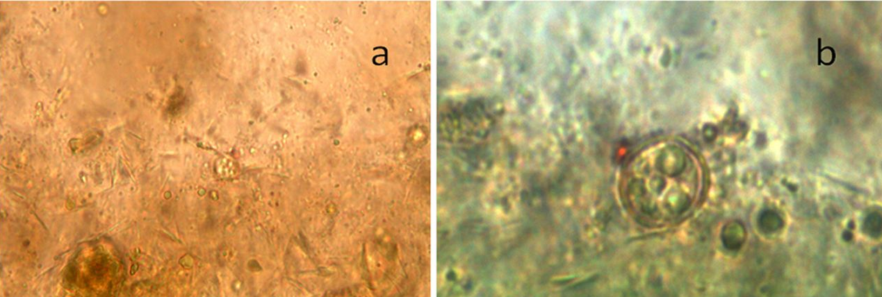
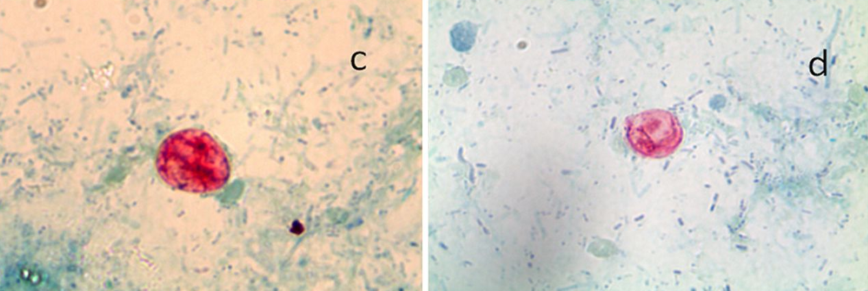
ชายอายุ 43 ปี ถ่ายอุจจาระเป็นน้ำมา 15 วัน


   ชายอายุ 43 ปี มาพบแพทย์เนื่องจากถ่ายอุจจาระเป็นน้ำมา 15 วัน ร่วมกับเบื่ออาหาร (น้ำหนักลดไป 3 กิโลกรัมใน 10 วัน) มีลมในท้องและปวดบิดเป็นครั้งคราว คลื่นไส้ และมีไข้ต่ำๆ. แพทย์ได้ส่งตรวจอุจจาระเพื่อหาพยาธิและไข่พยาธิ และเพาะเชื้อแบคทีเรีย ผลออกมาปกติ. เนื่องจากผู้ป่วยยังถ่ายอุจจาระเหลวอยู่ตลอด แพทย์จึงรับผู้ป่วยเข้าโรงพยาบาลเพื่อ investigation ต่อไป.
การตรวจร่างกายแรกรับ: อยู่ในเกณฑ์ปกติ ผู้ป่วยไม่มี signs ของ dehydration และไม่มี. evidence ของ systemic infection.

Lab: He had isolated monocytosis (1,300 cells/μl) with normal renal, hepatic, and pancreatic functions.
-Thyroid hormone and allergologic evaluations : no abnormality.
-The results for the classic parasitic serology examinations performed on travelers were negative, including the results for filariasis, strongyloidiasis, anisakiasis, ascariasis, distomatosis, trichinosis, toxocariasis, and bilharzia.
Gastroesophageal endoscopy : a peptic erosive esophagitis (level 2) unrelated to the symptoms.
-Small bowel biopsy specimen and a new series of three fresh stool samples were cultured for common enteropathogens (Salmonella, Shigella, Campylobacter, and Yersinia spp.) : negative results.
-Ova-and-parasite exam was โดยทำ concentration และ centrifuge stool specimen. The first microscopic examination involved direct wet smears with normal microscopy (magnifications, ×200 and ×400) (Fig. 1a) for detection of single-cell parasites and worms. Some structures were observed and were more evident following oil immersion (magnification, ×1,000) (Fig. 1b). Wet stool preparations viewed using a fluorescence microscope with UV illumination showed fluorescent spherical bodies.


Posted by : cpantip , E-mail : (chpantip@gmail.com) ,
Date : 2014-06-02 , Time : 10:02:33 , From IP : 172.29.3.129


ความคิดเห็นที่ : 1


   Modified acid-fast staining (Ziehl-Neelsen) was performed, and structures measuring 8 to 10 μm were observed microscopically (oil immersion; magnification, ×1,000) (Fig. 1c and d) in significant quantities.

1. การวินิจฉัยโรคคืออะไร
2. จะ manage อย่างไร


Posted by : cpantip , Date : 2014-06-02 , Time : 10:05:50 , From IP : 172.29.3.129

ความคิดเห็นที่ : 2


   ขอโทษค่ะ ลงรูปสลับกัน

Posted by : cpantip , Date : 2014-06-02 , Time : 10:06:57 , From IP : 172.29.3.129

ความคิดเห็นที่ : 3


   ขอตอบค่ะ

1. Diagnosis : Cyclosporiasis ค่ะ จากการพบ oocyst 8 to 10 μm ใน Modified acid-fast staining ของอุจจาระของผู้ป่วย
2. Management
-ส่งเลือดตรวจ HIV antibody
-ให้ยา co-trimoxazole ค่ะ


Posted by : June10 , Date : 2014-06-03 , Time : 13:29:36 , From IP : 172.29.3.129

ความคิดเห็นที่ : 4


   Diagnosis: Cyclospora cayetanensis infection.

Cyclospora cayetanensis เป็น intestinal coccidian protozoon ที่ปรากฎเป็นเชื้อที่ก่อโรคในคนทั่วโลก โดยทำให้เกิด self-limiting gastroenteritis ใน immunocompetent hosts. การแพร่กระจายของพยาธินี้จากคนสู่คนเกิดขึ้นโดยทางอ้อมจากการปนเปื้อนของ oocysts ในน้ำ อาหาร หรือดิน. พยาธินี้พบได้ทั่วโลก และมีถิ่นของโรคในเขต tropical และ subtropical. พบการติดเชื้อนี้ได้บ่อยในคนที่เดินทางไป Latin America, the Indian subcontinent และ Southeast Asia, รวมถึงจีน. ดังนั้น การติดเชื้อ Cyclospora cayetanensis จึงจัดอยู่ในกลุ่มของ traveler”s diarrhea.


Posted by : cpantip , Date : 2014-06-03 , Time : 15:16:24 , From IP : 172.29.3.129

ความคิดเห็นที่ : 5


   Cyclospora cayetanensis เป็นสาเหตุที่สำคัญของ waterborne และ food-borne outbreaks ที่เกิดจากผลไม้และผักที่นำเข้าสหรัฐอเมริกาจากประเทศกำลังพัฒนา. การที่มีรายงานโรคนี้น้อยกวาความเป็นจริงน่าจะเกิดจากการตรวจที่เป็นมาตรฐานหาไข่พยาธิและพยาธิไม่ได้รวมถึงการ identify Cyclospora oocysts. การที่มองข้ามโรคนี้ไปเนื่องจากผู้ที่ทำงานด้านสาธารณสุขและการแพทย์ยังไม่ตระหนักถึงโรคนี้.
หลักเกณฑ์ของการวินิจฉัย cyclosporiasis (โดย CDC ในปี 1997): ตรวจพบ oocysts ในอุจจาระ, intestinal fluid, หรือ small bowel biopsy specimens โดย microscopic examination; การพบ oocyst sporulation; และการตรวจพบโดย molecular methods (ซึ่งไม่ใช่ routine parasitology screening tool).

Oocysts ของCyclospora cayetanensis มีลักษณะกลม ขนาดเส้นผ่าศูนย์กลาง 8.0 - 10 μm, โดยมีเปลือกหนา 50 nm และมี outer threadlike coat ซึ่งผู้วิจัยบางรายเรียกว่า wrinkle (ดู Fig. 1a และ b). Diagnostic tests ยังมี phase-contrast microscopy, modified acid-fast staining ซึ่งจะติดสีแตกต่างกันไป (ตั้งแต่สีอ่อนๆ จนถึงแดง) (ดู Fig. 1c และ d), และ autofluorescence with UV illumination. ควรส่งตรวจอุจจาระหลายครั้งติดกันหลายวันเนื่องจากมี oocyst ถูกขับออกมาเป็นครั้งคราว. สำหรับผู้ป่วยรายนี้ พบ oocyst ในอุจจาระ 2 specimen จาก 3 specimen ที่ส่งตรวจ.

การรักษา Cyclospora infection คือการกิน trimethoprim-sulfamethoxazole 2x2 นาน 7 วัน ซึ่งได้ผลดีในผู้ป่วยรายนี้.

แพทย์ต้องนึกถึง Cyclospora infection ไว้ด้วยในผู้ป่วยที่มี chronic diarrhea.

Reference: Pons S, et al. Watery Diarrhea in an Immune-Competent Traveler.
J. Clin. Microbiol 2012 ; 50 : 3821-2.


Posted by : cpantip , Date : 2014-06-03 , Time : 15:19:10 , From IP : 172.29.3.129

สงวนสิทธิ์การแสดงความคิดเห็นสำหรับ สมาชิกเท่านั้น