ความคิดเห็นทั้งหมด : 3

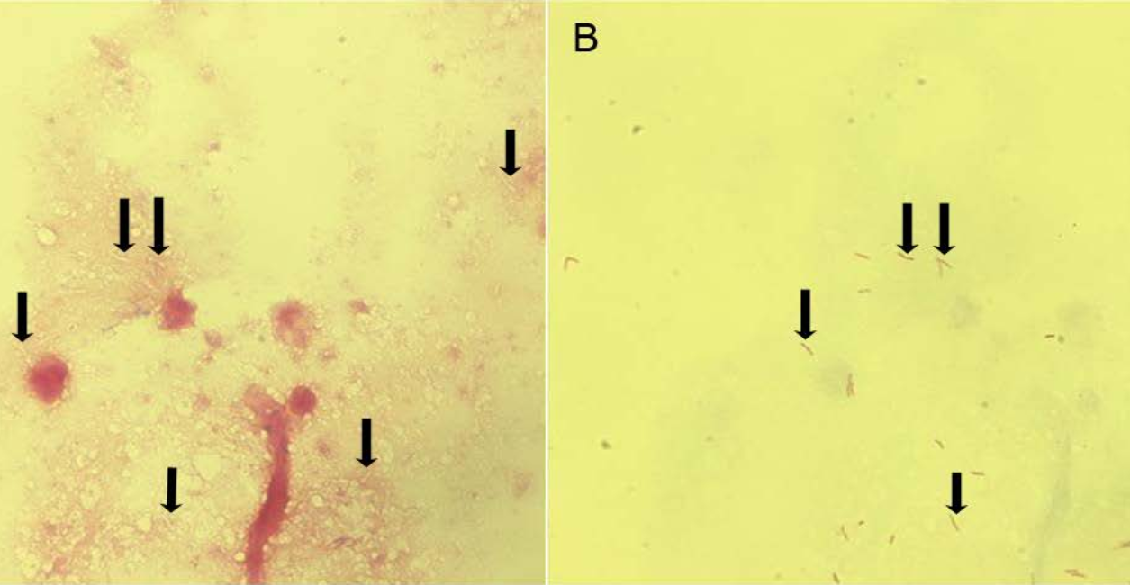
หญิงอายุ 32 ปี ไอ อ่อนเพลีย มีไข้กลางคืน และน้ำหนักลดมา 12 สัปดาห์.


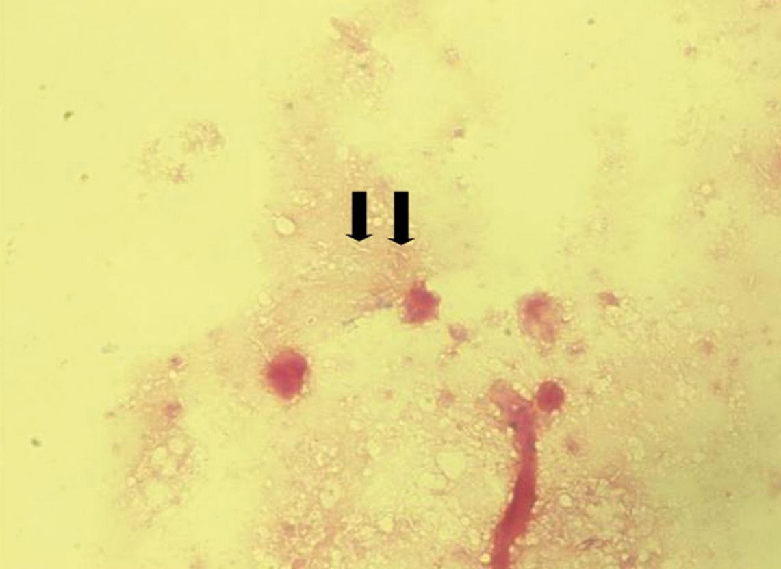
   หญิงอายุ 32 ปีมาพบแพทย์เนื่องจากไอมีเสมหะซึ่งบางครั้งมีเลือดปน อ่อนเพลีย มีไข้กลางคืนและน้ำหนักลดมา 12 สัปดาห์. ผู้ป่วยไม่สูบบุหรี่.

Physical examination revealed no significant abnormalities, and normal breath sounds were noted during auscultation.

-Blood analysis : hemoglobin concentration of 7.1 G%, white blood cell count of 15,100 cells/cu mm, and C-reactive protein concentration of 29 mg/liter.
-HIV antibody: negative.
-ส่งเสมหะเพื่อตรวจ microscopy และ bacterial + mycobacterial cultures.
ในรูปคือ Gram stain ของ sputum ของผู้ป่วย

1. การวินิจฉัยคืออะไร
2. จะ manage อย่างไร


Posted by : cpantip , E-mail : (chpantip@gmail.com) ,
Date : 2014-05-14 , Time : 11:11:52 , From IP : 172.29.3.141


ความคิดเห็นที่ : 1


   Problem list

1. Prolonged fever
2. Chronic productive cough + hemoptysis
3. Constitutional symptom
4. Anemia

คิดถึงว่ามี chronic pulmonary infection จาก mycobacterial infection ซึ่งนึกถึง M.tuberculosis มากที่สุด เพราะจาก clinical course. และ specimen ตรวจพบ gram ghost appearance bacilli

ส่งตรวจ sputum AFB CXR เพิ่มเติมก่อนครับ รวมทั้ง CBC ดู peripheral bloodsmear

การรักษาคงเป็น supportive ตามอาการ เฝ้าระวังภาวะ sepsis และ massive hemoptysis ยังไม่เริ่ม antiTB ครับ แต่ถ้า CXR แสดงลักษณะ apical upper lobe alveolar/interstitial infiltration ชัดเจนก็เริ่มยา antiTB เลยครับ

อาจจะต้องซักถามเรื่องปวดหัวเพิ่มเติมด้วยครับ เพราะเป็นมานานอาจมี tuberculous meningitis ร่วมด้วย

ส่วนเชื้ออื่นๆที่เห็น gram ghost ก็เป็น mycobacterium ตัวอื่นๆที่มี pulmonary involvement กลุ่ม m.kansassii, MAC คิดว่าอาการและ setting ยังไม่ค่อยเหมือนครับ

ส่วนเรื่องซีดถ้า non- massive hemoptysis แต่ออกมาเรื่อยๆคนไข้อาจจะซีดได้ แต่ก็แปลกตรงไม่น่าซีดขนาดนี้ ซีดขนาดนี้ น่าจะต้อง massive hemoptysis vital signs น่าจะไม่ดี ซึ่งปกติ TB มักจะไม่ค่อย massive hemoptysis ยกเว้นที่เป็น Rasmussen aneurysm แตก แต่ทั้งนี้คงต้อง work up cause of anemia ด้วย ระวัง mycobacterial involvement of bone marrow ครับ


Posted by : เป็ด , E-mail : (samadhi_66@hotmail.con) ,
Date : 2014-05-14 , Time : 13:42:45 , From IP : ppp-27-55-218-27.rev


ความคิดเห็นที่ : 2


   การวินิจฉัยโรค Pulmonary tuberculosis.

สิ่งที่ย้อมไม่ติดสีใน Gram stain ที่มีลูกศรชี่มีชื่อเรียกว่า ghost mycobacteria.
Cell wall ของ mycobacteria เป็น strongly hydrophobic เนื่องจากมี mycolic acids อยู่ ป้องกัน hydrophilic Gram stain reagents เข้าสู่เซลล์. ดังนั้น mycobacteria เป็น Gram neutral และสามารถปรากฎเป็น unstained structures หรือ Gram-positive beaded structures ใน the Gram stain.

สิ่งที่ต้องทำต่อไปคือย้อมสี Ziehl-Neelsen (ZN) ของ slide อันเดิมก็จะเห็น acid-fast bacilli ในจุดที่เป็น Gram neutrality ได้อย่างชัดเจน (ดูรูป 2).

chest radiograph ของผู้ป่วยพบว่ามี dense airspace opacities in the lingula and cavity formation in the left upper lobe ซึ่งเข้าได้กับ of pulmonary tuberculosis.
Sputum และ bronchoalveolar lavage fluid เพาะเชื้อขึ้น Mycobacterium tuberculosis.

ผู้ป่วยได้รับการรักษาหายด้วยการใช้ยา standard short-course chemotherapy.


Posted by : cpantip , Date : 2014-05-15 , Time : 13:43:07 , From IP : 172.29.3.141

ความคิดเห็นที่ : 3


   รูป 2 Gram and Ziehl-Neelsen stains of the sputum sample of the patient.
Panel A shows the Gram stain with slender linear, unstained structures (black arrows) in a background of Gram negative stained amorphous material and some degraded white blood cells.
Panel B is a Ziehl-Neelsen stain of the same smear spot and reveals that the Gram neutral structures are in fact acid-fast rods (black arrows). These proved to be M. tuberculosis.

Reference: van Ingen J, et al. Female with long-standing cough. Clin. Microbiol 2013 ; 51 :1065 and 1353.


Posted by : cpantip , Date : 2014-05-15 , Time : 14:02:27 , From IP : 172.29.3.141

สงวนสิทธิ์การแสดงความคิดเห็นสำหรับ สมาชิกเท่านั้น