ความคิดเห็นทั้งหมด : 7

A 55-YOM on hemodialysis had acute-onset LLQ abdominal pain for 12 h.


   ชายอายุ 55 ปีมาตรวจที่ห้องฉุกเฉินเนื่องจากปวดที่ท้องส่วนล่างข้างซ้ายแบบ acute onset มา 12 ชั่วโมง. ผู้ป่วยรับการทำ haemodialysis มา 20 ปีเนื่องจากมี diabetic nephropathy. ผู้ป่วยปวดน้อยลงเมื่อนอนหงายโดยงอเข่าข้างซ้าย

On examination, his temperature was 37.8°C and other vital signs were normal. The patient dragged his left leg when walking. The left lower quadrant of the abdomen was tender and the psoas sign (pain on hip extension) was positive on the left.

CBC: white cell count ปกติ, แต่ C-reactive protein สูงเล็กน้อย. แพทย์นึกถึง psoas abscess แต่ enhanced CT และ plain MRI ไม่พบรอยโรคใดๆ.

1. การวินิจฉัยน่าจะเป็นอะไร
2. จะ manage อย่างไร


Posted by : cpantip , E-mail : (chpantip@medicine.psu.ac.th) ,
Date : 2014-01-31 , Time : 13:00:11 , From IP : 172.29.3.164


ความคิดเห็นที่ : 1


   ชายอายุ 55 ปี
1. ปวดที่ท้องส่วนล่างข้างซ้ายแบบ acute onset มา 12 ชั่วโมง
มี psoas sign (pain on hip extension) was positive on the left.
2. ESRD on hemodialysis

คิดว่าน่าจะเป็น Lt psoas abscess มากที่สุดค่ะ
การวินิจฉัยแยกโรค - hematoma?
Plan: 1. hemoculture แล้วให้ empiric antibiotic ไปเลย เนื่องจากเป็นผู้ป่วย hospital acquired setting เชื้อก่อโรคน่าจะเป็น Staphylococcus aureus มากที่สุด แต่ก็ควรรักษาครอบคลุม MRSA ไปเลย ส่วนเชื้ออันดับรองคือ E. coli คิดว่าจะให้ยา Vancomycin+ceftriaxone รอผล H/C เพื่อปรับยา
2. ขอทำ enhanced CT scan ซ้ำ แต่ห่างสัก 2 วันเพราะผู้ป่วยเพิ่งเป็นมา 12 ชม. image study อาจยังไม่เห็น


Posted by : daisy1 , Date : 2014-02-02 , Time : 15:13:11 , From IP : 172.29.3.164

ความคิดเห็นที่ : 2


   คุณหมอ Daisy1 ตอบถูกแล้วค่ะ

แต่เนื่องจากไม่มีการวินิจฉัยอื่นๆที่เข้ากับอาการโรคผู้ป่วยรายนี้มากกว่า psoas abscess และเขาก็มี risk factor หลายอย่างสำหรับ psoas abscess. ดังนั้น แพทย์ผู้รักษาจึงให้ provisional diagnosis เป็น early imaging-negative psoas abscess (inflammatory process ก่อน overt abscess formation).


Posted by : cpantip , Date : 2014-02-03 , Time : 12:21:05 , From IP : 172.29.3.164

ความคิดเห็นที่ : 3


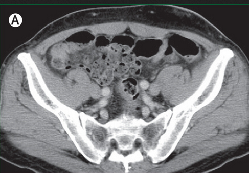
   Imaging-negative psoas abscess
(A) No abnormal findings on enhanced CT on day of admission.


Posted by : cpantip , Date : 2014-02-03 , Time : 12:22:42 , From IP : 172.29.3.164
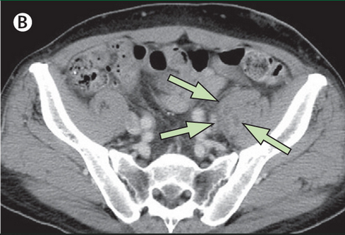
ความคิดเห็นที่ : 4


   (B) Repeat enhanced CT on day 3 after admission shows multiple hypodense foci with ring enhancement in the left psoas muscle (arrows).

ผู้ป่วยได้เข้ารับการรักษาในโรงพยาบาลและได้รับการรักษาด้วย IV vancomycin (500 mg every 48 h) หลังเจาะเลือดส่งเพาะเชื้อไป 2 ขวด. วันที่ 3 ของการอยู่โรงพยาบาล ได้รับการทำ enhanced CT ซ้ำซึ่งพบ multiple hypodense foci with ring enhancement ที่ left psoas muscle, ซึ่งยืนยันการวินิจฉัย psoas abscess (figure).

ผลการเพาะเชื้อของเลือดขึ้น grew methicillin-sensitive Staphylococcus aureus.

ถาม จะเปลี่ยนยาปฏิชีวนะหรือไม่ และจะ investigate อะไรต่อ


Posted by : cpantip , Date : 2014-02-03 , Time : 12:23:58 , From IP : 172.29.3.164

ความคิดเห็นที่ : 5


   เปลี่ยนค่ะ เปลี่ยนจาก vancomycin เป็น IV cloxacillin หรือ cefazolin เพราะ มี activity ต่อ MSSA ดีกว่า vancomycin ซึ่งเป็น weak bactericidal drug.

Investigation เพื่อหา source ของ MSSA bacteremia
คือ ทำ echocardiography เพื่อหาว่ามี infective endocarditis หรือไม่ ค่ะ


Posted by : daisy1 , Date : 2014-02-03 , Time : 14:26:58 , From IP : 172.29.3.164

ความคิดเห็นที่ : 6


   แพทย์ได้ส่ง transoesophageal echocardiography ซึ่งไม่พบว่ามี vegetation หรือintracardiac abscess. ดังนั้น source ของ infection น่าจะเป็น arteriovenous fistula ของผู้ป่วย. ผู้ป่วยตอบสนองต่อการรักษาด้วย IV cefazolin (2 กรัมหลัง dialysis สัปดาห์ละ 3 ครั้ง นาน 6 สัปดาห์). การติดตามที่ 2 เดือนหลังจากนั้น ผู้ป่วยสบายดี ไม่มี relapse.

Posted by : cpantip , Date : 2014-02-04 , Time : 11:39:23 , From IP : 172.29.3.164

ความคิดเห็นที่ : 7


   Psoas abscess พบได้น้อย แม้ว่าจะสามารถตรวจพบได้ง่ายยิ่งขึ้นจากการใช้ imaging studies. แบ่ง psoas abscess ได้เป็น primary และ secondary. Primary psoas abscess เกิดขึ้นจาก hematogenous หรือ lymphatic spread ของการติดเชื้อจากตำแหน่งที่อยู่ไกล ขณะที่ secondary psoas abscess เกิดจาก direct extension ของการติดเชื้อจากตำแหน่งที่อยู่ใกล้ๆ.
เชื้อก่อโรคของ primary abscess ที่พบบ่อยที่สุดคือ Staphylococcus aureus, ขณะที่ mixed enteric flora พบได้บ่อยใน secondary psoas abscess.
ปัจจัยเสี่ยงของ primary abscess คือ เบาหวาน, intravenous drug use, HIV infection, renal failure, และสาเหตุอื่นๆ ของ immunosuppression. มีรายงานผู้ป่วยจำนวนมากที่มี primary psoas abscess ในผู้ป่วยที่ทำ dialysis เนื่องจากมี hemodialysis access septicemia.
อาการนำของ psoas abscess มีหลากหลาย ตั้งแต่ที่เป็น classic triad คือ fever, back pain, and limp (ไข้ ปวดหลัง และเดินกระโผลกกระเผลก) ซึ่งพบเพียง 30% ของผู้ป่วย. การทำ hyperextension ของ hip ทำให้ปวดเนื่องจากไปยืด psoas muscle ที่มีฝี. CT scan โดยทั่วไปแล้วให้ definite diagnosis ได้ดีมากและจัดเป็น gold standard แม้ว่าผู้เชี่ยวชาญบางท่านรายงานว่า MRI ไวกว่า CT. ในผู้ป่วยรายนี้ ทั้ง CT และ MRI ในระยะแรกไม่พบความผิดปกติทั้งคู่ ซึ่งแสดงว่า psoas abscess ในระยะต้นๆ อาจตรวจไม่พบด้วยวิธีเหล่านี้. ก่อนหน้านี้มีรายงานผู้ป่วยรายหนึ่งซึ่งพบว่ามีข้อจำกัดของ plain CT สำหรับการวินิจฉัย early psoas abscess.
การรักษา psoas abscess ต้องการการเริ่มให้ยาปฏิชีวนะทันทีและการระบายหนอง. อัตราตายของ primary psoas abscess คือ 2.4%, แต่สูงขึ้นเป็น 18.9% ใน secondary abscess และสูงถึง 100% ในรายที่ไม่ได้รับการรักษา. สาเหตุของการตายที่สำคัญคือ การรักษาช้าหรือรักษาไม่เพียงพอ. ดังนั้น การวินิจฉัยที่ถูกต้องและเริ่มต้นการรักษาที่เหมาะสมอย่างรวดเร็วจึงมีความสำคัญมาก. ถ้าคิดว่าผู้ป่วยเป็น early psoas abscess ก็ควรเริ่มต้นการรักษาด้วยยาปฏิชีวนะทันที แม้ว่า imaging studies จะได้ผลลบก็ตาม.

Reference: Takada T, et al. Imaging-negative psoas abscess. Lancet 2014;383:280.


Posted by : cpantip , Date : 2014-02-06 , Time : 13:59:37 , From IP : 172.29.3.164

สงวนสิทธิ์การแสดงความคิดเห็นสำหรับ สมาชิกเท่านั้น