A 29-year-old soldier had a two-day history of headache and feverทหารอายุ 29 ปี มาด้วยอาการไข้และปวดหัวมากมา 2 วัน เขามีน้ำใสๆไหลออกมาจากรูจมูกข้างซ้ายมา 2 ปี หลังจากเกิดอุบัติเหตุรถจักรยานยนต์ 1. ท่านคิดว่าผู้ป่วยปวดหัวและมีไข้จากอะไร 2. ท่านจะตรวจร่างกายหาอะไรเพื่อการวินิจฉัย 3. ท่านจะ investigate อะไรบ้าง (เท่าที่จำเป็น) 4. จะให้ empirical therapy ด้วยยาอะไร Posted by : chpantip , E-mail : (chpantip@medicine.psu.ac.th) , Date : 2008-06-27 , Time : 08:15:05 , From IP : 172.29.3.68 |
1. Traumatic CSF leakage จากมี fracture base of skull ค่ะ 2. การครวจร่างกาย - วัด V/S เพื่อดู signs of Increase ICP และ SIRS - ตรวจ Neurological signs; GCS, pupil RAPD Facial palsy nystagmus facial sensetion (CN V, VI, VII) ตรวจการได้ยินม ตรวจ carotid atery เพื่อหา pseudoaneurysm และ caroticocavernous fistula 3. คิดว่าจำเป็นต้อง Ct scan เพื่อหาตำแหน่ง fracture และส่ง CSF for cell count ceff diff, protein-sugar, gram stain, culture และส่งตรวจ Electrolyte เพื่อดู SIDH complication 4. การรักษา - Empirical Antibiotic; Ceftriaxone หรือ cefotaxime ค่ะ - Surgical repair or lumbar drainage->> post traumat 7 วัน ยังปิดเองไม่ได้ Posted by : nongnham , E-mail : (nongnham025@gmail.com) , Date : 2008-06-28 , Time : 16:01:55 , From IP : 222.123.181.211 |
ลืมตอบข้อ 1 ค่ะ สาเหตุการปวดศีรษะและมีไข้เกิดจาก Meningitis --> น่าจะเป็น Bacterial meningitis มากที่สุดค่ะ Posted by : nongnham , E-mail : (nongnham025@gmail.com) , Date : 2008-06-28 , Time : 16:05:20 , From IP : 222.123.181.211 |
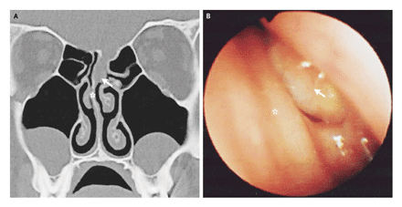
ขอบคุณคุณหมอ Nongnham มากค่ะ สำหรับคำตอบ ที่ตอบมาถูกต้องแล้วค่ะ ผู้ป่วยมี CSF rhinorrhea หลังอุบัติเหตุก็ต้องนึกว่าเขามี fractue base of skull หรือ bony defect ทีไหน ในรูป A คือ Coronal computed tomography B คือ Endoscopic rhinoscopy เขามี acute onset ของอากรไข้และปวดหัว ก็ต้องนึกถึง acute bacterial meningitis คำถาม 1. เชื้อก่อโรคในผู้ป่วยรายนี้น่าจะเป็นเชื้ออะไรคะ 2. จาก CT scan of brain และ endoscopic rhinoscopy มีความผิดปกติอะไรบ้าง Posted by : chpantip , E-mail : (chpantip@medicine.psu.ac.th) , Date : 2008-06-30 , Time : 08:44:00 , From IP : 172.29.3.68 |
1. เชื้อที่เป็นสาเหตุคิดว่าเป็น Streptococcus pneumoniae มากที่สุดค่ะ รองลงมาคือ H.influenzae ค่ะ 2. CT brain; haziness of Lt ethmoid sinus extended to both frontal sinus (ลูกศรชี้) , Lt inferior and middle conchae โตกว่าด้านขวา และใน Lt etmoid sinus มีลักษณะเป็น mass อยู่ข้างใน (ข้างๆลูกศร) Rhinoscopy; swelling of inferior conchae และมี mass ยื่นลงมาเหนื่อ inferior conchae (ลูกศรชี้) Posted by : nongnham , E-mail : (nongnham025@gmail.com) , Date : 2008-06-30 , Time : 12:13:08 , From IP : 222.123.142.191 |
คุณหมอ Nongnham ตอบถูกแล้ว เก่งมากค่ะ ขอเล่า progression ต่อนะคะ Lumbar puncture: white-cell count = 4100/cumm, 98 % เป็น neutrophil ซึ่งเป็นลักษณะของ bacterial meningitis, ผลเพาะเชื้อ CSF ไม่ขึ้น . Coronal computed tomography: absence of the left ethmoidal plate and a focal brain herniation (Panel A, arrow). Incidental maxillary-sinus retention cysts were seen bilaterally. Endoscopic rhinoscopy: a small, pulsating mass consistent with the presence of an encephalocele (Panel B, arrow). The asterisks indicate the mid-septum. ผู้ป่วยรายนี้มี fistulous defect ในnasal cavity มีผลทำให้ CSF รั่วออกมา และมี meningitis ตามมา ผู้ป่วยได้รับการผ่าตัด corrective surgery โดยตัด encephalocele, ตามมาด้วยปิด bony defect. ได้ติดตามผู้ป่วยไปนาน 2 ปี ผู้ป่วยแข็งแรงดี ไม่เกิด meningitis อีกเลย สำหรับเชื้อก่อโรคใน setting ของ bacterial meningitis ตามหลัง CSF rhinorrhea ที่พบมากที่สุดคือ Streptococcus pneumoniae รองมาคือ Haemophilus influenzae ค่ะ Posted by : chpantip , E-mail : (chpantip@medicine.psu.ac.th) , Date : 2008-07-01 , Time : 08:50:28 , From IP : 172.29.3.68 |