ความคิดเห็นทั้งหมด : 10

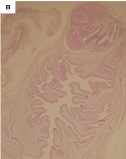
A 30 YOM with multiple subcutaneous nodules


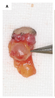
   ชายอายุ 30 ปี คลำได้ก้อนใต้ผิวหนังที่คอด้านซ้าย ผู้ป่วยไม่มีไข้ ไม่เจ็บที่ก้อน ต่อมามีก้อนลักษณะเดียวกันทะยอยขึ้นมามากกว่า 10 ก้อน ที่หนังตาล่าง ด้านข้างของจมูก คาง และหน้าท้อง.
แพทย์ตรวจพบว่ามี subcutaneous nodules จึงทำ biopsy ได้ชิ้นเนื้อดังรูป

1. การวินิจฉัยคืออะไร
2. จะ manage อย่างไร


Posted by : cpantip , E-mail : (chpantip@medicine.psu.ac.th) ,
Date : 2013-07-19 , Time : 12:22:48 , From IP : 172.29.3.187


ความคิดเห็นที่ : 1


   1. คิดว่าผู้ป่วยเป็นโรค cysticercosis ค่ะ
2. รอ confirm ด้วย histopathology ของ subcutaneous nodule ซึ่งจะพบว่ามี scolex with hooks
เมื่อยืนยันแล้วว่ามี cysticercosis ก็ต้องหาว่ามี cysticerci ที่อวัยวะที่เป็นอันตราย คือ สมอง และตา หรือไม่ เพราการรักษาด้วยยา ฆ่าพยาธิคือ praziquantel และ albendazole ทำให้เกิดอันตรายต่อสมองและตาได้จากปฏิกิริยาของร่างกายต่อพยาธิที่กำลังตาย


Posted by : lara , Date : 2013-07-20 , Time : 15:25:48 , From IP : 172.29.3.187

ความคิดเห็นที่ : 2


   คุณหมอ Lara ตอบถูกแล้วค่ะ ขอถามเพิ่มเติมนะคะ
-ผู้ป่วยเป็นโรคนี้ได้อย่างไรคะ


Posted by : cpantip , Date : 2013-07-23 , Time : 13:37:36 , From IP : 172.29.3.187

ความคิดเห็นที่ : 3


   เป็น cysticercosis จากการกินไข่พยาธิ (Taenia egg) ค่ะ ไข่พยาธินี้อาจปนเปื้อนในอาหารประเภทผักดิบจากการเอาอุจจาระไปทำปุ๋ยรดผัก หรือไข่พยาธิอาจติดอยู่ที่มือของคนทำอาหารแล้วไปปนเปื้อนในอาหาร

Posted by : lara , Date : 2013-07-23 , Time : 14:11:54 , From IP : 172.29.3.187

ความคิดเห็นที่ : 4


   คุณหมอ Lara ตอบถูกแล้วค่ะ

ผู้ป่วยรายนี้เป็นชาวญี่ปุ่น ครั้งแรกที่พบว่ามีก้อนที่คอด้านซ้าย คือ ธค. 2009. ต่อมาพบว่ามีก้อนที่อื่นขึ้นมาอีก. มีปล้องพยาธิออกมากับอุจจาระเมื่อเดือนเมย. 2010. ผู้ป่วยได้ไปพบแพทย์เมื่อเดือนมิย 2010. ประวัติที่ได้คือ ผู้ป่วยไปอินเดีย 3 ครั้งในช่วงเวลา สค.2008 ถึง เมย 2009 โดยพักอยู่ที่อินเดียครั้งละ 1 เดือน. ตอนอยู่ที่อินเดีย ผู้ป่วยพักอยู่กับครอบครัวชาวอินเดียและกินอาหารพื้นบ้าน ซึ่งมักเป็นหมูย่าง และผักสด ไม่เคยกินเนื้อสัตว์ดิบๆ เลย

Physical examination showed that more than 10 subcutaneous lesions in total were palpable in the left inferior eyelid, left wing of the nose, lower jaw, neck, and left lateral abdomen areas.

Skin biopsy ที่ลำตัว: subcutaneous white round-shaped cyst is a viable cysticercus (รูป1A).

Histopathology : Cysticercus section showing an invaginated neck, and scolex with two suckers and hooklet (H-E stain, ×40). (รูป 1B).


Posted by : cpantip , Date : 2013-07-24 , Time : 11:34:49 , From IP : 172.29.3.187

ความคิดเห็นที่ : 5


   Computed tomography scan จากคอไปถึง pelvis.
-Cysticerci were found in (A) each cheek, (B) the lower jaw, (C and E) chest and abdominal walls, and (D and F) the lower back. There were more than 20 cysticerci spread throughout the body of our patient. (รูป 2).


Posted by : cpantip , Date : 2013-07-24 , Time : 11:36:09 , From IP : 172.29.3.187

ความคิดเห็นที่ : 6


   Magnetic resonance images of the brain showed 30 small cysts within the parenchyma, 4–8 mm in diameter, most of which were associated with ring enhancement and some with peri-lesion focal edema (รูป 3A),
ผู้ป่วยไม่มีอาการทางระบบประสาทใดๆ.


Posted by : cpantip , Date : 2013-07-24 , Time : 11:37:38 , From IP : 172.29.3.187

ความคิดเห็นที่ : 7


   นอกเหนือจากการตรวจพบทาง histopathology และ imaging study แพทย์ได้ส่ง serological test โดยใช้ T. solium cysticercus-specific antigens ด้วย immunoblots ได้ผลว่าผู้ป่วยเป็น cysticercosis. ผู้ป่วยให้ประวัติว่ามีปล้องเหมือนเส้นหมี่ออกมาทางทวารหนักหลายครั้ง แพทย์จึงให้การรักษา taeniasis ด้วย Gastrographin แต่ไม่มีพยาธิถูกขับออกมา. จากการตรวจอุจจาระ พบ taeniid eggs และการทำ copro-polymerase chain reaction (PCR)และ copro-loop-mediated isothermal amplification (LAMP) พบว่าเป็น Indian haplotype ของ T. solium โดย mitochondrial DNA analysis (GenBank accession no.: AB066489).

ดังนั้น ผู้ป่วยจึงได้รับการวินิจฉัยเป็น disseminated cysticercosis ร่วมกับมี taeniasis.


Posted by : cpantip , Date : 2013-07-24 , Time : 11:41:25 , From IP : 172.29.3.187

ความคิดเห็นที่ : 8


   ผู้ป่วยได้รับการรักษาด้วย albendazole 400 มก.วันละ 2 ครั้งนาน 28 วันร่วมกับ prednisolone 1 มก/กก/วัน สำหรับ disseminated cysticercosis และยากันชักคือ valproate sodium. ผู้ป่วยมีไข้ ปวดหัว และกดเจ็บที่ subcutaneous nodules ในวันที่ 3 หลังเริ่มการรักษา และอาการเหล่านี้ก็หายไปในวันเดียวกัน. ในระหว่างที่ได้รับยา subcutaneous nodules ค่อยๆ หายไป และ จาก magnetic resonance images ของสมอง พบว่าเมื่อสิ้นสุดการรักษา parenchymal lesions หายไปหมด(รูป 3B). ระดับของ antibody ก็ค่อยๆ ลดลงไปได้วยระหว่างการรักษา. จากการติดตาม ผู้ป่วยไม่มีอาการใดๆ นานกว่า 6 เดือน.

Posted by : cpantip , Date : 2013-07-25 , Time : 11:20:22 , From IP : 172.29.3.187

ความคิดเห็นที่ : 9


   ผลของ serological analyses โดยใช้ Taenia solium-specific antigens. (A) Immunoblot analysis โดยใช้ serum samples ก่อนและหลังการรักษาด้วย albendazole ขนาด 400 มกวันละ 2ครั้งนาน 28 วัน. Probe แต่ละ strip ด้วย individual serum sample ที่ 1:20 dilution.
Lane N, negative control; lane P, positive control;
lane 1, before treatment, lane 2, Day 0 before start of chemotherapy; lane 3, Day 28 after start of chemotherapy; lane 4, Day 30 after stopping chemotherapy; lane 5, Day 90 after stopping chemotherapy. Serum sample reacting antigens ระหว่าง 10 to 30 kDa เป็น serologically diagnosed as cysticercosis.


Posted by : cpantip , Date : 2013-07-25 , Time : 11:24:22 , From IP : 172.29.3.187

ความคิดเห็นที่ : 10


   (B) Kinetics of anti-T. solium-specific antigens antibody levels obtained by using enzyme-linked immunosorbent assay (ELISA). Start of chemotherapy is at Day 0.

Posted by : cpantip , Date : 2013-07-25 , Time : 11:25:38 , From IP : 172.29.3.187

สงวนสิทธิ์การแสดงความคิดเห็นสำหรับ สมาชิกเท่านั้น