ความคิดเห็นทั้งหมด : 7

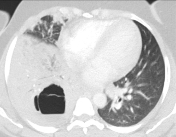
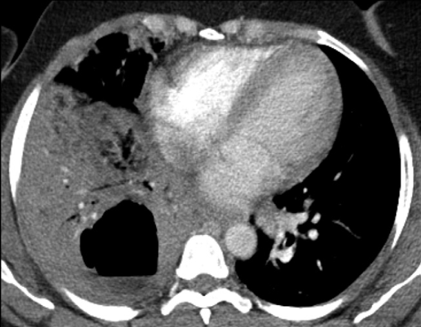
หญิง 37 ปี มี cavitary lung lesion with worsening eosinophilia


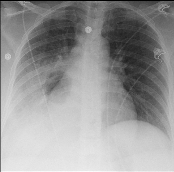
   หญิงอายุ 37 ปีมีไข้ไอหอบมา 2 วัน ผู้ป่วยได้รับการวินิจฉัย community-acquired pneumonia และได้เข้ารับการรักษาในโรงพยาบาล. ภาพรังสีทรวงอกพบright lower lobe consolidation. ผู้ป่วยแข็งแรงดีมาก่อน.
CBC: WBC 9,500/cumm with neutrophil 91%, eosinophil 2%. ผู้ป่วยตอบสนองดีต่อการรักษาด้วย levofloxacin และได้กลับบ้าน. 4 วันต่อมา ผู้ป่วยต้องเข้ารับการรักษาในโรงพบยาบาลอีกเนื่องจากมีไข้ ไอ หอบอีก.
On physical examination, the patient had a blood pressure of 124/73 mm Hg, a pulse rate of 110 beats per minute, a respiratory rate of 40 breaths per minute, and a temperature of 39° C. Her oxygen saturation was 100% on room air. Decreased breath sounds and egophony were noted at the right lung base.
CBC: WBC 13,000/cumm with a neutrophil 65%and eosinophil 9%.

Chest x-ray: persistent right lower-lobe consolidation with a new air lucency (รูป 1).


Posted by : cpantip , E-mail : (chpantip@medicine.psu.ac.th) ,
Date : 2013-04-11 , Time : 13:44:34 , From IP : 172.29.3.93


ความคิดเห็นที่ : 1


   Chest computed tomography (CT) showed a cavity measuring 6 × 5 × 7 cm within the right lower-lobe consolidation and a floating membrane inside the cavity (รูป 2 และ 3).

Posted by : cpantip , Date : 2013-04-11 , Time : 13:47:03 , From IP : 172.29.3.93

ความคิดเห็นที่ : 2


   Chest computed tomography (CT) showed a cavity measuring 6 × 5 × 7 cm within the right lower-lobe consolidation and a floating membrane inside the cavity (รูป 2 และ 3).

On bronchoscopy: The tracheal and right main-stem bronchial mucosa had an irregular cobblestone appearance ; purulent secretions were evident in the right lower-lobe bronchus. Mucosal biopsy of the carina revealed eosinophilic infiltrates without additional diagnostic features. Cytologic and microbial smears (bacteria, fungi, mycobacteria) of bronchial washings were negative.

อีก 5 วันต่อมาผู้ป่วยมี peripheral eosinophilia สูงขึ้นเป็น 7,620/cumm (47% eosinophils).

1. การวินิจฉัยน่าจะเป็นโรคใด


Posted by : cpantip , Date : 2013-04-11 , Time : 13:48:58 , From IP : 172.29.3.93

ความคิดเห็นที่ : 3


   จากการที่ผู้ป่วยมี increasing eosinophilia คิดว่าผู้ป่วยมี parasitic infection ที่จะเป็น lung cavity และมี floating membrane อยู่ใน cavity จาก CT scan คือ hydatid cyst ค่ะ


Posted by : lara , Date : 2013-04-11 , Time : 15:35:37 , From IP : 172.29.3.93

ความคิดเห็นที่ : 4


   คุณหมอ Lara ตอบถูกแล้วค่ะ
การวินิจฉัยแยกโรค cavitating lung lesion ร่วมกับมี peripheral eosinophilia คือ
1. Infection
1.1 bacterial (eg, brucellosis),
1.2 fungal (eg, histoplasmosis, coccidioidomycosis, และ aspergillosis),
1.3 parasitic (eg, schistosomiasis, paragonimiasis, และ strongyloidiasis)
2. vasculitis หรือ autoimmune diseases (eg, Churg-Strauss syndrome และgranulomatosis with polyangiitis);
3. eosinophilic pneumonias; และ
4. drug reactions
ดู Table 1.

ผล serum autoantibodies และ vasculitis panel ได้ผลลบ.
stool examination ได้ผลลบสำหรับ parasitic ova, larvae และ protozoa.


Posted by : cpantip , Date : 2013-04-14 , Time : 14:40:18 , From IP : 172.29.3.93

ความคิดเห็นที่ : 5


   wet preparation ของ sputum พบ echinococcal hooklets ( รูป 4).

การส่ง serum echinococcal antibodies IgE และ IgG ได้ผลบวก สนับสนุนว่าผู้ป่วยเป็น, pulmonary cystic echinococcosis.


Posted by : cpantip , Date : 2013-04-16 , Time : 14:53:36 , From IP : 172.29.3.93

ความคิดเห็นที่ : 6


   Echinococcosis เป็น zoonotic infection ที่เกิดจาก tapeworms (cestodes) ใน genus Echinococcus. Human echinococcosis มี 3 รูปแบบ คือ cystic, alveolar และ polycystic. Cystic และ alveolar echinococcosis มีความสำคัญเพราะพบได้ทั่วโลก ขณะที่ polycystic echinococcosis พบได้เฉพาะที่ Central และ South America.

ผู้ป่วยรายนี้มี cystic echinococcosis ที่เกิดจาก E. granulosus, ซึ่ง endemic ที่ใน Northern และ Eastern Africa, Southern Europe, Western China, Australia และ South America. สัตว์กินเนื้อ (carnivores) เช่น สุนัข หมาป่า และหมาจิ้งจอกเป็น definitive host สำหรับพยาธินี้. คนเป็นโรคนี้จากการกินไข่ของ Echinococcus ที่ถูกขับออกมากับ feces ของ infected carnivores. หลังกินไข่พยาธินี้เข้าไป larva ถูกปลดปล่อยออกมา ไชผ่านผนังลำไส้ และเดินทางไปสู่อวัยวะต่างๆ หรือในช่องท้อง แล้วเกิดเป้น hydatid cyst ซึ่งค่อยๆ โตขึ้น สร้าง protoscolices (immature tapeworms) และ daughter cysts อยู่ภายใน cyst. ไม่มี mature tapeworms เกิดขึ้นในคน. ตำแหน่งที่พบ hydatid cyst ได้บ่อย คือ ตับ (69-75%), ปอด (17-22%), ไต(1-4%), และ intraabdominal cavity (2-5%).

Alveolar echinococcosis เกิดจาก E. multilocularis, ซึ่งส่วนใหญ่พบที่ตับเท่านั้น E. multilocularis แทรกซึมในอวัยวะ ในลักษณะคล้ายเนื้องอก เกิดเป็น vesicle เล็กๆ จำนวนมาก. ส่วน polycystic echinococcosis, เกิดจาก E. vogeli หรือ E. oligarthrus,มีลักษณะเป็น cyst จำนวนมากที่มีหลายขนาด (multiple cysts of various sizes) พบได้ที่ตับ ปอด และม้าม. โดยทั่วไปแล้ว alveolar และ polycystic echinococcoses ทำให้เกิดอาการโรครุนแรงกว่า cystic infection.

เนื่องจาก cysts โตขึ้นช้าๆ, ผู้ป่วยส่วนใหญ่ที่มี cystic echinococcosis จะไม่มีอาการใดๆ ในช่วงแรกของโรค. ต่อมา อาการของผู้ป่วยขึ้นอยู่กับอวัยวะที่เป็นโรค และขนาดของ cysts. ผู้ป่วยที่มี pulmonary cystic echinococcosis มักมาพบแพทย์ด้วยเรื่องไข้ ไอ หอบเหนื่อย และเจ็บหน้าอก ส่วน hepatic echinococcosis ผู้ป่วยมักมาด้วยตับโตและ right epigastric pain


Posted by : cpantip , Date : 2013-04-16 , Time : 14:56:50 , From IP : 172.29.3.93

ความคิดเห็นที่ : 7


   Ultrasonography และ CT มีประโยชน์ในการตรวจหา hepatic lesions ขณะที่ chest x-ray และ CT ช่วยในตรวจหา pulmonary cavitary หรือ cystic lesions. ถ้า cyst ที่แตกเชื่อมต่อกับ tracheobronchial tree การระบาย contents ใน cyst ออกไปทำให้เห็นเป็น air-fluid level. หลังจากมีการขับ cystic fluid ออกไปบางส่วน อาจมองเห็น cystic membranes ที่ ยุบตัวลงได้ (ซึ่งมีชื่อเรียกว่า serpent sign). ในผู้ป่วยรายนี้ พบลักษณะดังกล่าวใน CT scan ก่อนจะทำ bronchoscopy ซึ่งแสดงว่าผู้ป่วยมี partially ruptured hydatid cyst (รูป 3). ถ้า cyst ยุบลงหมด endocyst ที่ยู่ยี่ลอยอยู่บนน้ำที่อยู่ใน cavitary lesion ทำให้เกิดเป็น water-lily sign. ถ้า sign ดังกล่าว เกือบจะจำเพาะต่อ pulmonary echinococcosis ซึ่งต้องพิจารณาประกอบกับอาการโรคด้วย

ไม่มี laboratory test อันใดอันหนึ่งที่เป็น diagnostic test สำหรับ cystic echinococcosis. Peripheral eosinophilia พบเพียง 15 - 25% ของผู้ป่วยโรคนี้ โดยเกิดขึ้นเมื่อ cyst ที่แตกปล่อย parasite antigens เข้าสู่กระแสเลือด. ในผู้ป่วยรายนี้ cyst rupture ทำให้ peripheral eosinophilia สูงขึ้น และพบ echinococcal hooklets ในเสมหะ. Cyst rupture สามารถทำให้ผู้ป่วยเสียชีวิตได้จากการชักนำให้เกิด anaphylactic shock, respiratory distress, severe asthma-like symptoms, และ sepsis.


Posted by : cpantip , Date : 2013-04-24 , Time : 11:48:14 , From IP : 172.29.3.216

สงวนสิทธิ์การแสดงความคิดเห็นสำหรับ สมาชิกเท่านั้น