ความคิดเห็นทั้งหมด : 8

A 38 YOM had painful penile edema & urethral blood discharge after anal SI.


   ชายอายุ 38 ปี มาพบแพทย์เนื่องจากมีอาการบวมที่อวัยวะเพศและหนังหุ้ม ที่อวัยวะเพศบวมแข็งๆ ปวดอยู่ตลอดเวลา มีน้ำเลือดออกมาจากท่อปัสสาวะ และมีก้อนเลือดปนออกมากับปัสสาวะ. อาการเหล่านี้เกิดขึ้นหลังการร่วมเพศกับชายคนหนึ่ง.
มีประวัติสูบบุหรี่และดื่มเหล้ามานาน

1. การวินิจฉัยน่าจะเป็นอะไร
2. จะ manage อย่างไร


Posted by : cpantip , E-mail : (chpantip@medicine.psu.ac.th) ,
Date : 2013-03-04 , Time : 10:40:02 , From IP : 172.29.3.93


ความคิดเห็นที่ : 1


   ชายอายุ 38 ปี
Problems
1. บวมที่อวัยวะเพศและหนังหุ้ม ที่อวัยวะเพศบวมแข็งๆ ปวดอยู่ตลอดเวลา
2.มีน้ำเลือดออกมาจากท่อปัสสาวะ และมีก้อนเลือดปนออกมากับปัสสาวะ. อากาiเกิดขึ้นหลังการร่วมเพศกับชายคนหนึ่ง (anal intercourse).
3.สูบบุหรี่และดื่มเหล้ามานาน

เขามี STD มี การอักเสบของ penis และ urethra คิดถึงการติดเชื้อ
1. Neisseria gonorrhoeae
2. Chlamydia trachomatis
3. Herpes simplex virus (HSV)
4. Treponema pallidum (the causative agent of syphilis)
5. Haemophilus ducreyi (the causative agent of chancroid)
7. Chlamydia trachomatis serovars L1-3 (the causative agents of lymphogranuloma venereum; LGV)
8. Klebsiella granulomatis (the causative agent of Granuloma inguinale; also known as “Donovanosis”)
เขาน่าจะมีแผลใน urethra ถ้ามี painful ulcers นึกถึง HSV และ chancroid มากกว่า ส่วน ulcers ของ syphilis, LGV และ granuloma inguinale ทั่วไปแล้วไม่เจ็บ.


management
1. investigation
■HIV antibody
■Urethral or urine test (culture or nucleic acid amplification) for gonorrhea and chlamydia.
■Syphilis serology
2. การรักษา ให้ยา ceftriaxone IV และ doxycycline กิน


Posted by : lara , Date : 2013-03-05 , Time : 16:02:01 , From IP : 172.29.1.74

ความคิดเห็นที่ : 2


   ขอบคุณคุณหมอ Lara สำหรับคำตอบค่ะ

ผู้ป่วยได้ไปตรวจที่โรงพยาบาล และได้รับยา levofloxacin 500 mg IV ทุก 24 ชม.นาน 10 วัน; ceftriaxone 1 g IV วันละ 2 ครั้งนาน 5 วัน และกิน clindamycin 300 mg x 4 นาน 10 วัน. หลังได้รับยาไป 3 วันตำแหน่งที่แข็งๆ ที่ prepuce แตกออกมาเองเป็นหนองปนเลือดซึ่งไม่มีกลิ่น. ผู้ป่วยไม่มีไข้และไม่มีอาการผิดปกติอื่นๆ. ผู้ป่วยได้กลับไปรพ.อีก

The clinical examination showed localized dermatosis affecting the trunk, the genital region including the penis at its shaft and the distal portion of the glans, the scrotum and the pubis. Dermatosis was characterized by a painful and progressively growing ulcer with an erythematous base and irregular borders. Additionally, the ulcer was necrotic in appearance, was covered by a fibroid material, and produced a discharge. Deformation of the penis was evident, giving the impression of a circumcised penis.

ผู้ป่วยได้รับการผ่าตัด debridement และ biopsy lesion.
ผล routine clinical laboratory tests อยูในเกณฑ์ปกติทั้งหมด. ผู้ป่วยได้รับ cefuroxime (750 mg IV, every 8 hr) นาน 7 วันแล้วแพทย์ให้ผู้ป่วยกลับบ้าน. ผล biopsy : balanitis with acute and chronic inflammatory cell infiltration, multiple pyogenic microabscesses, and the absence of neoplastic cells.


Posted by : cpantip , Date : 2013-03-06 , Time : 11:40:33 , From IP : 172.29.3.93

ความคิดเห็นที่ : 3


   ในระยะเวลา 2 สัปดาห์ รอยโรคใหญ่ขึ้นและผู้ป่วยได้กลับไปรพ.อีก และได้เข้ารับการรักษาในรพ.ในแผนก Urology.

The physical examination showed severe erosion of the prepuce skin and areas of extended necrosis. The posterior side of the penis showed a large area of skin erosion with serous and purulent material. There was an open abscess in the suprapubic area draining an odorless, bloody, and purulent material, and the scrotum was also affected, with multiple ulcers (รูป 1).


Posted by : cpantip , Date : 2013-03-06 , Time : 11:42:15 , From IP : 172.29.3.93

ความคิดเห็นที่ : 4


   (A) Macroscopic lesions in patient 1. A view of the dorsal region of the penis showing a large ulcerative lesion with irregular borders. Part of the ulcer is covered with a fibrinoid discharge. Anterior view of the genital region showing the ulcerative lesion on the pubis, an ulcerative lesion on the penis with necrotic lesions at the borders and the total absence of the prepuce.

Posted by : cpantip , Date : 2013-03-06 , Time : 11:49:07 , From IP : 172.29.3.93

ความคิดเห็นที่ : 5


   ถึงตอนนี้ หน่วยผิวหนังได้รับการปรึกษา และคิดถึงว่าผู้ป่วยอาจมีการติดเชื้อ E. histolytica. ผู้ป่วยได้รับการตรวจ
1. หาระดับของ serum anti-amoebic antibodies โดย enzyme-linked immunosorbent assay (ELISA) technique ซึ่งได้ผลบวกอย่างชัดเจน (optical density [OD] at 490 nm = 0.60, cut-off point OD at 490 nm = 0.525), และ
2. microscopic detection of E. histolytica/E. dispar in the biopsy specimen (รูป1B)
3. polymerase chain reaction (PCR) characterization of Entamoeba species extracted from the environmental DNA obtained from the purulent material of the damaged tissue.


Posted by : cpantip , Date : 2013-03-06 , Time : 11:52:27 , From IP : 172.29.3.93

ความคิดเห็นที่ : 6


   (B) Microphotography of the biopsy specimen obtained after 10 days of metronidazole treatment. The tissue slides were stained with periodic acid-Schiff (PAS). An infiltration of mononuclear inflammatory cells and the presence of red stained trophozoites of Entamoeba histolytica/Entamoeba dispar were observed.

Posted by : cpantip , Date : 2013-03-06 , Time : 11:53:10 , From IP : 172.29.3.93

ความคิดเห็นที่ : 7


   หลังจากการวินิจฉัย cutaneous amebiasis, ผู้ป่วยได้รับการรักษาด้วย metronidazole (500 mg IV, every 8 hr) และ ceftriaxone (1 g IV, every 12 hr). หลังจากได้รับยาทั้งสองนาน 30 วัน ฝีหาย และมี granulation tissue เป็นบริเวณกว้างและไม่มีผิวหนังปกคลุมรวมทั้งที่ penis ทั้งหมด. หลังจากนั้น ผู้ป่วยต้องได้รับการผ่าตัด reconstructive surgery และทำ autologous skin transplant.

Posted by : cpantip , Date : 2013-03-06 , Time : 11:54:08 , From IP : 172.29.3.93

ความคิดเห็นที่ : 8


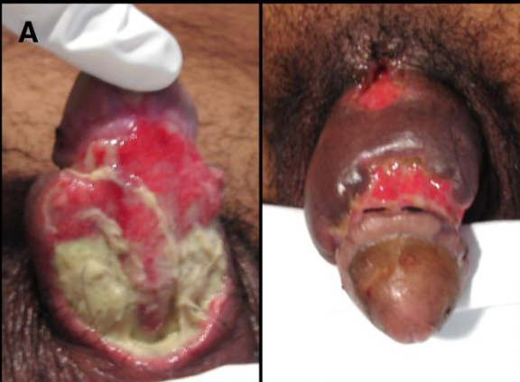
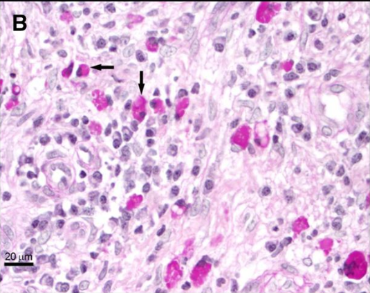
   มีรายงาน cutaneous amebiasis ครั้งแรกเมื่อปี 1892 โดย Nasse รายงานผู้ป่วย amoebic liver abscess ซึ่งมีภาวะแทรกซ้อนหลังการระบายหนอง เกิดมี extensive ulceration และ necrosis ของ abdominal skin, subcutaneous tissue และ muscles. cutaneous amebiasis เป็น clinical form ที่พบน้อยที่สุดของ human amebiasis และโดยทั่วไป เป็นผลเนื่องมาจากการติดเชื้อ Entamoeba ของผิวหนังที่มีความเสียหายมาก่อน. Ngai และ Frazier ทบทวนรายงานที่ตีพิมพ์ก่อนปี 1936 พบว่ามีรายงาน 27 รายจากจีน, Indochina, และสหรัฐอเมริกา. หลังจากนั้น จำนวนผู้ป่วย cutaneous amebiasis ที่รายงานลดลงเป็น 11 รายในปี 1941. Magaña และคณะรายงาน 26 documented cases ของ cutaneous amebiasis จาก clinical files ของ Pediatric Dermatology Department of the General Hospital of Mexico of the Health Ministry, และจาก Pathology Department ของ Hospital of Medical Specialties of the Mexican Institute of Social Security (IMSS). อย่างไรก็ตาม ส่วนใหญ่ของผู้ป่วยรายงานในช่วงปี 1969 - late 1980s. ผู้ป่วยเหล่านี้มี deep skin lesions ที่ anal, perineal, vulval, vaginal และ genital regions, เช่นเดียวกับ diaper area ของทารกที่มีท้องเสียจาก Entamoeba histolytica. ในผู้ใหญ่ ตำแหน่งของ cutaneous amebiasis ที่พบบ่อยคือ abdominal area ซึ่งเกิดจาก involvement ของ abdominal wall ระหว่างที่มี spontaneous amoebic liver abscess หรือเป็นภาวะแทรกซ้อนจากการระบายหนอง amoebic liver abscess. การติดต่ออีกทางหนึ่งของ cutaneous amebiasis คือการร่วมเพศ.

รายนี้แสดงถึง sexually transmitted genital cutaneous amebiasis และเน้นความสำคัญของ clinical form นี้ ของ amoebic disease ซึ่งแม้ว่าจะพบ cutaneous amebiasis ได้น้อย แต่ก็ต้องเป็น differential diagnosis ของ ulcerative lesions ของ human genitalia, โดยเฉพาะอย่างยิ่งใน amebiasis-endemic areas และใน high risk groups. การวินิจฉัยให้ทันการ และการรักษาให้ตรงโรคมีความสำคัญในการป้องกัน serious anatomical damage และรอยโรคหายโดยไม่มี functional sequelae.

Ref: Morán P, et al. Cutaneous amebiasis: the importance of molecular diagnosis of an emerging parasitic disease.Am J Trop Med Hyg 2013;88: 186-190.


Posted by : cpantip , Date : 2013-03-07 , Time : 15:26:00 , From IP : 172.29.3.93

สงวนสิทธิ์การแสดงความคิดเห็นสำหรับ สมาชิกเท่านั้น