ความคิดเห็นทั้งหมด : 3

ชาย 79 ปี เหนื่อยหอบมา 8 ชั่วโมง และมี abnormal CXR


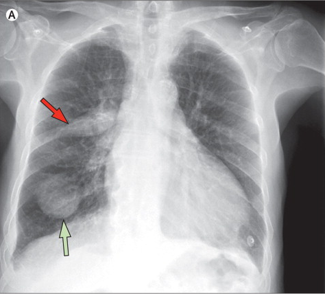
   ชายอายุ 79 ปีมารพ.เนื่องจากเหนื่อยหอบมา 8 ชั่วโมงและมีขาบวมทั้งสองข้าง. ผู้ป่วยมี hypertension, atrial fibrillation และ chronic obstructive pulmonary lung disease. เขาได้รับยา ipratropium bromide, irbesartan, amlodipine, amiodarone และ furosemide มานาน.

He had normal vital signs and the only relevant findings in physical examination were bilateral edema of both legs and rales in the lower half of both lungs.

ในรูป A คือ CXR ของผู้ป่วย ทำเมื่อแรกรับ
1. พบความผิดปกติอะไร การวินิจฉัยน่าจะเป็นอะไร
2. จะ manage อย่างไร


Posted by : cpantip , E-mail : (chpantip@medicine.psu.ac.th) ,
Date : 2013-01-14 , Time : 15:56:53 , From IP : 172.29.3.93


ความคิดเห็นที่ : 1


   ชายอายุ 79 ปี
Problem 1. Congestive heart failure (เหนื่อยหอบมา 8 ชั่วโมง มีขาบวมทั้งสองข้าง, rales in the lower half of both lungs).
2. Underlying diseases: hypertension, atrial fibrillation และ chronic obstructive pulmonary lung disease
3. Abnormal CXR: พบ Cardiomegaly, pulmonary congestion และมี lung mass อยู่ 2 ก้อนที่ปอดข้างขวา อันแรก ที่ RLL ลักษณะเป็นก้อนกลม ขอบเรียบ (ซึ่งน่าจะเป็น tumor หรืออาจเป็น round pneumonia ก็ได้ เพียงแต่ไม่มีประวัติ ไข้และไอ) ส่วนอีกก้อน รูปร่างคล้ายกระสวย อยู่ต่อเนื่องกับ right hilum และน่าจะเป็น fluid ใน minor fissure

คิดว่าผู้ป่วยมี left heart failure เป็นเรื่องสำคัญที่ต้องรีบให้การรักษา โดยให้ O2 และให้ IV furosemide และทำ CXR ซ้ำในวันรุ่งขึ้น ถ้า mass ยังอยู่ก็ควรทำ CT chest
สาเหตุของ acute left heart failure อาจเป็นจาก acute myocardial infarction ควรทำ ECG ค่ะ


Posted by : lara , Date : 2013-01-16 , Time : 11:16:11 , From IP : 172.29.3.93

ความคิดเห็นที่ : 2


   CXR: an opaque elliptic image (red arrow) in the horizontal fissure and a round tumor-like image in the right inferior lobe (green arrow) (รูปA).

ผู้ป่วยได้รับการรักษาด้วย furosemide แล้วผู้ป่วยมีอาการดีขึ้น.


Posted by : cpantip , Date : 2013-01-16 , Time : 14:29:09 , From IP : 172.29.3.93

ความคิดเห็นที่ : 3


   ได้ทำ CXR ซ้ำ 4 ชั่วโมงต่อมา พบว่า elliptic image หายไปและ round tumor-like image หายไปจนเกือบหมด. สองวันต่อมา หลังจากการปรับขนาดยา แล้ว ได้ทำ CXR อีกครั้ง ซึ่งพบว่าปกติ. (รูป B).

Phantom tumours (หรือ vanishing tumours) ของปอด เป็นผลจากการมี pleural effusion loculate อยู่ภายใน interlobar fissure ที่เกิดจาก exacerbated congestive heart failure. ไม่มีการรักษาอื่นๆ นอกเหนือจากการแก้ไข underlying heart condition และ volume overload.

การนึกถึง vanishing tumours มีความสำคัญ เพราะทำให้หลีกเลี่ยงการทำ diagnostic tests และการรักษาที่ไม่จำเป็นได้.

Reference: Oliveira E, et al. Phantom tumour of the lung. Lancet 2012;380:2028.


Posted by : cpantip , Date : 2013-01-16 , Time : 14:32:28 , From IP : 172.29.3.93

สงวนสิทธิ์การแสดงความคิดเห็นสำหรับ สมาชิกเท่านั้น