ความคิดเห็นทั้งหมด : 4

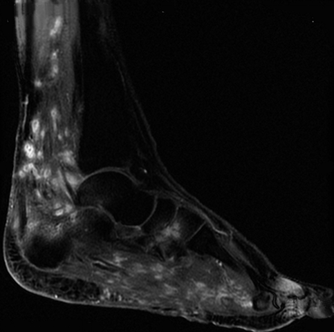
A 32-YOW had progressive swelling of the left foot for 6-years.


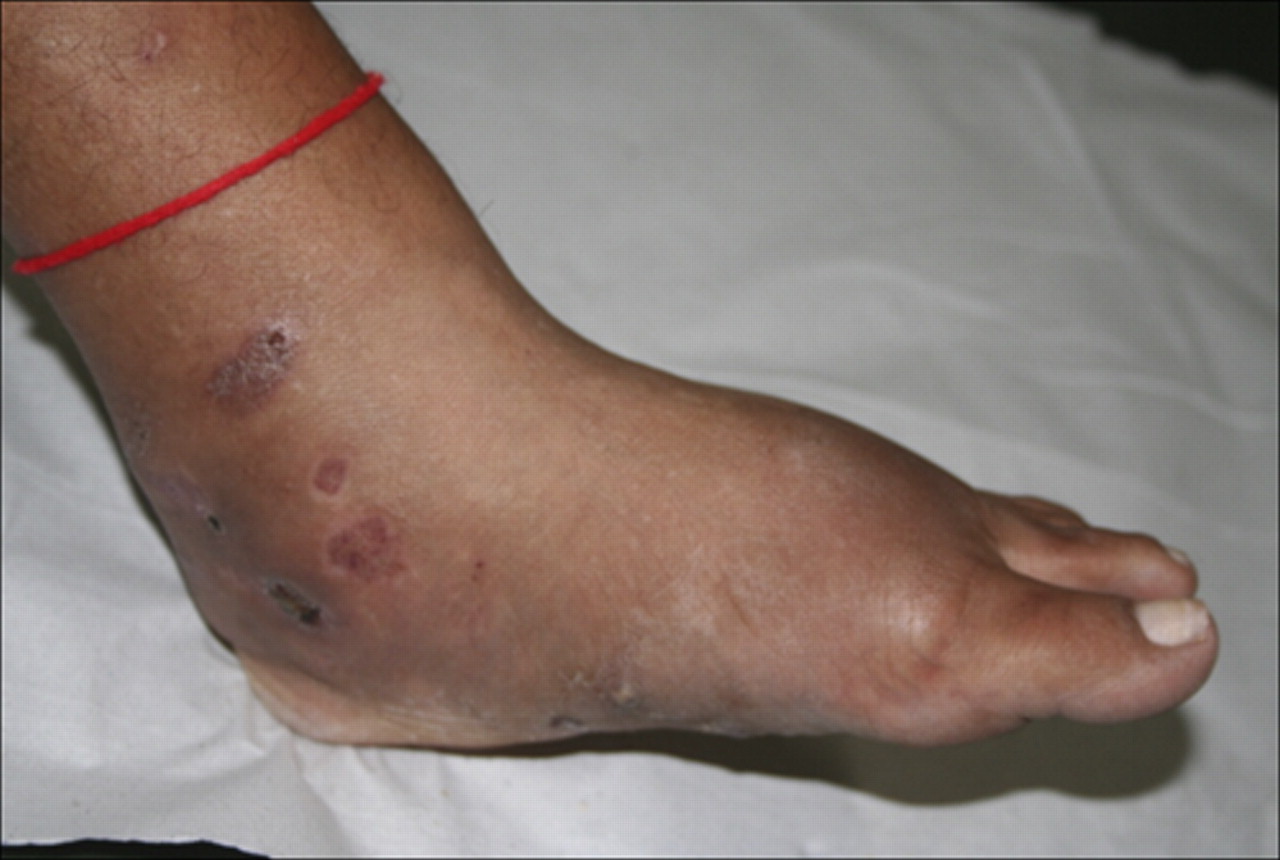
   หญิงอายุ 32 ปี อาชีพทำนา มารพ.เนื่องจากมีเท้าซ้ายบวมมากขึ้นมานาน 6 ปี. มีอาการบวมและแตกออกมาเป็นหนองร่วมกับมีเม็ดขาวๆ ออกมาด้วย (รูป 1).

1. การวินิจฉัยคืออะไร
2. จะ manage อย่างไร


Posted by : cpantip , E-mail : (chpantip@medicine.psu.ac.th) ,
Date : 2012-04-14 , Time : 14:40:09 , From IP : 172.29.3.14


ความคิดเห็นที่ : 1


   หญิงอายุ 32 ปี
ปัญหา
1. มีเท้าซ้ายบวมและผิดรูปมากขึ้นมานาน 6 ปี
2. Deformed foot with draining sinuses and seropurulent discharge
with white grains

จากลักษณะของเท้าที่ผิดรูปและมี sinuses และมี grain (ซึ่งเป็นการที่เชื้อมาจับกันเป็นก้อน) เข้าได้กับการวินิจฉัย mycetoma ซึ่งแยกเป็น eumycetoma (เกิดจากเชื้อรา) และ actinomycetoma (เกิดจาก Actinomycetes ซึ่งอาจเป็น Nocardia spp. หรือ Actinomyces ก็ได้)

การวินิจฉัยเชื้อ: ต้องเอา grain (หรือ granule) มาย้อม Gram stain และ modified acid fast stain, ทำ biopsy รอยโรค และส่งเพาะเชื้อ fungi, aerobic and anaerobic bacteria แล้วจึงให้การรักษาที่เฉพาะค่ะ


Posted by : lara , Date : 2012-04-16 , Time : 09:51:49 , From IP : 172.29.3.14

ความคิดเห็นที่ : 2


   คุณหมอ Lara ตอบถูกแล้วค่ะ

Clinical appearance ของsinus tracks ที่ medial aspect ของ ข้อเท้าขวาและข้อเท้าและเท้าบวมทั่วๆไป เข้าได้กับ mycetoma.

ได้ทำ Potassium hydroxide preparation ของหนอง พบว่ามี white-yellowish nodules with fine filaments. เมื่อนำเม็ดในหนองมาเพาะเชื้อบน Sabouraud agar และ casein hydrolysis test ยืนยันว่าเป็นเชื้อ Nocardia brasiliensis.
Hematoxylin/eosin-stained sections : microabscesses, granulomatous reaction, and nodules containing basophilic bacterial filaments peripherally surrounded by eosinophilic pillars.

สรุปว่า การวินิจฉัยทางคลินิกคือ mycetoma และได้รับการยืนยันว่าเป็น mycetoma จาก Nocardia brasiliensis.


Posted by : cpantip , Date : 2012-04-18 , Time : 08:18:23 , From IP : 172.29.3.14

ความคิดเห็นที่ : 3


   รูป 2 คือ Magnetic resonance imaging ของ right lower leg (sagittal section, post-gadolinium) : small nodules with intensive ring enhancement distributed in the distal left leg and foot along with bone marrow infiltration of the calcaneus. Some spherical lesions showed hyperintense signal in the periphery and low-signal intensity central foci consistent with the recently described "dot-in-circle sign".

Posted by : cpantip , Date : 2012-04-18 , Time : 10:34:23 , From IP : 172.29.3.14

ความคิดเห็นที่ : 4


   ผู้ป่วยได้รับการรักษาด้วย amikacin (ขนาดยา 15 มก/กก./วัน นาน 21 วัน ทั้งหมด 5 cycles), trimethoprim sulfamethoxazole (800/160 mg, b.i.d., นาน 2 ปี), และทำ debridement procedures 3 ครั้ง. การรักษาได้ผลดี.
Magnetic resonance imaging อาจมีประโยชน์ในการวินิจฉัย mycetoma ตั้งแต่ระยะต้น. พบได้บ่อยว่ามีการวินิจฉัยภาวะนี้ช้าเนื่องจากมีความจำกัดในการตรวจทางจุลชีววิทยาซึ่งทำให้เกิดภาวะแทรกซ้อนตามมาโดยอาจรุนแรงถึงขนาดต้องตัดขาผู้ป่วยได้.

Reference: Cordeiro F, et al. Mycetoma. Am J Trop Med Hyg 2011;85:791.


Posted by : cpantip , Date : 2012-04-18 , Time : 10:39:30 , From IP : 172.29.3.14

สงวนสิทธิ์การแสดงความคิดเห็นสำหรับ สมาชิกเท่านั้น