ความคิดเห็นทั้งหมด : 6

A 60-YOW had worsening nausea, vomiting & abdominal for 1 mo.


   หญิงอายุ 60 ปี มารพ.เนื่องจากคลื่นไส้อาเจียนและปวดท้องมากขึ้นมา 1 เดือน.
ผู้ป่วยเคยได้รับการวินิจฉัย suspected Crohn disease และได้รับ prednisone 20 มก/วันติดต่อกันมานานกว่า 10 ปี.

At presentation, the patient was afebrile. Her blood pressure
was 115/78 mm Hg, and her pulse was 126 beats/minute. She was alert and oriented. Her only complaint was generalized abdominal pain. Diffuse expiratory wheezes were heard on pulmonary examination. Her abdomen was distended, and she had tenderness upon palpation in the right upper quadrant. No rebound tenderness was noted. Bowel sounds were audible. The remainder of her physical exam was unremarkable.

Initial laboratory results:
CBC: WBC 8,500 cells/mm3 (86% neutrophils, 12% lymphocytes, 1% monocytes, and 1% eosinophils). Hb 12.4 gm/dL, and platelet count 366,000/mm3.

Serum lactate, 1.1 mg/dL; blood urea nitrogen, 48mg/dL; and serum creatinine, 0.60 mg/dL.

Liver function tests: total bilirubin at 0.6 mg/dL, and the serum
albumin level was low at 2.5 gm/dL.

Human immunode-ficiency virus (HIV) antibody assay was nonreactive.

Blood cultures remained negative. The patient was started on levo-
floxacin and metronidazole.

On hospital day 2, a computed tomography (CT) scan of her abdomen revealed edema of the right colonic wall and air in the wall of the cecum.
A chest radiograph disclosed patchy infiltrative changes in both lower lobes and consolidative changes in the right middle lobe. Infiltrates in the right middle and right lower lobes also were evident on a CT scan of the chest.

1. การวินิจฉัยที่น่าจะเป็นไปได้คืออะไร
2. จะ manage อย่างไรต่อไป


Posted by : cpantip , E-mail : (chpantip@medicine.psu.ac.th) ,
Date : 2012-04-04 , Time : 11:40:43 , From IP : 172.29.3.14


ความคิดเห็นที่ : 1


   ขอตอบค่ะ
หญิงอายุ 60 ปี
Problems
1. คลื่นไส้อาเจียนและปวดท้องมากขึ้นมา 1 เดือน.
-BP 115/78 mm Hg, P 126/min,
-tenderness at RUQ
-CT abdomen: edema of the right colonic wall and air in the wall of the cecum.
2. suspected Crohn disease ได้รับ prednisone 20 มก/วันติดต่อกันมานานกว่า 10 ปี.
4. Lungs: Diffuse expiratory wheezes
-CXR + CT chest: patchy infiltration at both lower lobes and right middle lobe.

ผู้ป่วยมีพยาธิสภาพที่ปอดและสำไส้ใหญ่ และเป็น immunocompromised host เนื่องจากกิน prednisolone มาเป็นเวลานาน สาเหตุของโรคน่าจะเป็นการติดเชื้อแบบ subacute คือ
1. TB:: pulmonary + intestinal
2. Parasites นึกถึง hyperinfection syndrome ของ Strongyloides stercoralis มากที่สุด แม้จะไม่มี eosinophilia ก็เป็นได้
Investigation ต่อไป ควรทำ
1. stool exam + sputum exam for larva ของ Strongyloides stercoralis
2. colonoscopy
3. sputum for AFB+culture for TB

Treatment: ผู้ป่วยมี pulse เร็ว (P 126/min),ต้องหาสาเหตุและประเมินเรื่อง volume depletion แล้วแก้ไข
พบ air in the wall of the cecum จาก CT abdomen (pneumatosis intestinalis) ซึ่งอาจเป็นอาการแสดงของโรคที่รุนแรงหลายอย่างซึ้งได้แก่ gastrointestinal infection, inflammatory bowel disease, life-threatening intraabdominal catastrophe (เช่น bowel infarction หรือมี underlying malignant condition) ต้องพิจารณาเรื่อง surgical treatment ด้วยค่ะ


Posted by : lara , Date : 2012-04-07 , Time : 13:54:37 , From IP : 172.29.3.14

ความคิดเห็นที่ : 2


   คุณหมอ lara ตอบถูกแล้วค่ะ

ผู้ป่วยได้รับการผ่าตัด exploratory laparotomy with a subsequent right-sided hemicolectomy ในวันที่ 3 ของการอยู่รพ. ในการผ่าตัดพบascites, lymphadenopathy along the ileocolic vessels, and thickening of the cecum and descending colon.
ผลของ pathological examination ของ colon แสดงในรูป 1.


Posted by : cpantip , Date : 2012-04-08 , Time : 14:11:58 , From IP : 172.29.3.14

ความคิดเห็นที่ : 3


   รูป A: Chronic active colitis with Strongyloides was visualized within a colonic crypt (arrow);

รูป B, Mucosal ulceration with invasive Strongyloides (arrow) was noted in the colon;


Posted by : cpantip , Date : 2012-04-08 , Time : 14:16:38 , From IP : 172.29.3.14

ความคิดเห็นที่ : 4


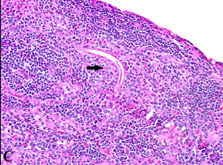
   รูป C, Strongyloides involvement was evident in this lymph node from the colonic mesentery (arrow);

Posted by : cpantip , Date : 2012-04-08 , Time : 14:17:38 , From IP : 172.29.3.14

ความคิดเห็นที่ : 5


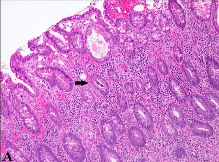
   รูป D; Cytopathological examination ของ colon ยังพบความผิดปกติอื่นอีกด้วยดังในรูปนี้

การวินิจฉัยในรูป D คืออะไร


Posted by : cpantip , Date : 2012-04-08 , Time : 14:19:58 , From IP : 172.29.3.14

ความคิดเห็นที่ : 6


   พบ endothelial cell ที่ตัวโตและมี intranuclear inclusion body ค่ะ
การวินิจฉัย CMV co-infection with disseminated strongyloidiasis (มี lymph node involvement ด้วยค่ะ)

การรักษา disseminated strongyloidiasis ให้ ivermectin+albendazole
CMV colitis ให้ gancyclovir ค่ะ


Posted by : lara , Date : 2012-04-11 , Time : 15:17:36 , From IP : 172.29.3.14

สงวนสิทธิ์การแสดงความคิดเห็นสำหรับ สมาชิกเท่านั้น