ความคิดเห็นทั้งหมด : 7

A 32-YOM with pemphigus vulgaris had multiple discrete, punched-out ulcers for 2 wks.


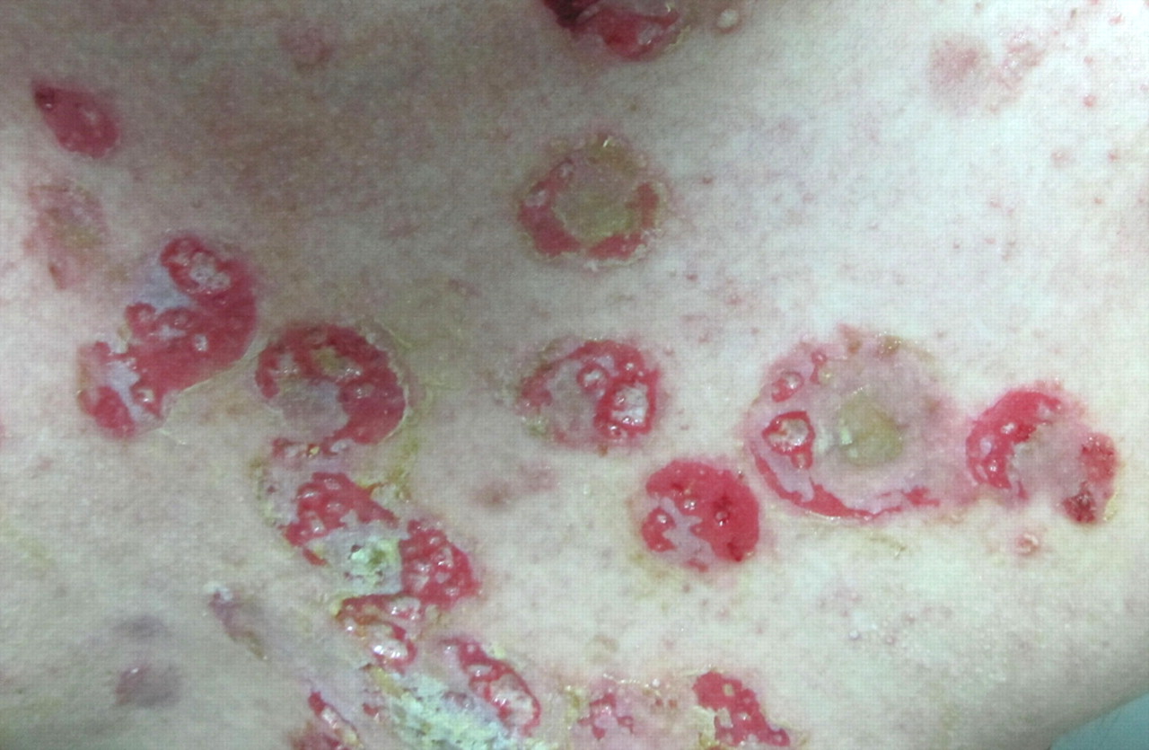
   ชายอายุ 32 ปี
เมื่อ 2 เดือนก่อน ผู้ป่วยมีถุงน้ำ (blister) กระจายทั่วตัว. การวินิจฉัย pemphigus vulgaris ได้รับการยืนยันโดย pathological examination และ direct immunofluorescence assays ของ skin biopsy specimen และโดย indirect immunofluorescence assays ของ serum samples. ในเบื้องต้น ผู้ป่วยได้รับการรักษาด้วย oral prednisolone (30–40 มก/วันนาน 11 วัน) แต่ไม่ตอบสนองต่อการรักษา แพทย์จึงเพิ่มขนาดของ prednisolone เป็น 90 มก/วันซึ่งก็มีการตอบสนองเพียงเล้กน้อย. ผู้ป่วยจึงได้รับ prednisolone ขนาดสูงขึ้นเป็น 180 มก/วัน ซึ่งทำให้รอยโรคส่วนใหญ่เริ่มมี reepithelialize.
อย่างไรก็ตาม 2 สัปดาห์ต่อมา ผุ้ป่วยเริ่มมีแผลจำนวนมากเกิดขึ้นที่รอยแผลเดิมของ pemphigus. ผู้ป่วยไม่มีความผิดปกติอื่นๆ.
Physical examination revealed many rice grain–sized, discrete, punchedout ulcers covered with yellowish, moist necrotic debris on the
chest, upper back, and abdomen (รูป 1). Some of the ulcers
subsequently became confluent.


Posted by : cpantip , E-mail : (chpantip@medicine.psu.ac.th) ,
Date : 2012-02-23 , Time : 12:52:15 , From IP : 172.29.3.14


ความคิดเห็นที่ : 1


   ได้ทำ incisional biopsy รอยโรคที่หน้าอก และได้ส่ง specimen เพื่อ pathological examination (รูป 2, รูป B คือรูปขยายของ A)
และส่ง bacterial, mycobacterial และ fungal culture.

1. การวินิจฉัยคืออะไร
2. จะ manage อย่างไร


Posted by : cpantip , Date : 2012-02-23 , Time : 13:04:34 , From IP : 172.29.3.14

ความคิดเห็นที่ : 2


   Patient : Male 32 years old
Chief complaint : Multiple discreted punched – out ulcer for 2 weeks
Problem lists :
1. Multiple discreted punched – out ulcer for 14 day.
2. Pemphigus vulgaris for 60 day.
3. High dose steroid exposure.
History discussion : 1.Where is the localizing of the pathology? จากประวัติของผู้ป่วยที่มีพยาธิสภาพเด่นที่บริเวณผิวหนัง ที่มีลักษณะกระจัดกระจายทั่วร่างกายโดยที่เมื่อดูประกอบกับการตรวจร่างกายแล้วพบว่าเป็น"แผลตื้นๆที่ปรกคลุมโดยเนื้อตายซีดๆ"นั้นสื่อถึงการที่พยาธิสภาพทำให้เกิดเนื้อตายผ่านกระบวณการลดเลือดไปยังบริเวณดังกล่าว ทำให้การระบุตำแหน่งของพยาธิสภาพจากบริเวณผิวหนังชั้นบนๆได้แคบและจำเพาะลงมาอีกว่าจะต้องเป็นที่บริเวณของหลอดเลือดเล็กๆในบริเวณดังกล่าว 2.What is the causes of the localized pathology? สำหรับตำแหน่งของพยาธิสภาพที่กล่าวไว้ว่า มันจะต้องอยู่ที่หลอดเลือดบริเวณผิวหนัง ในที่นี้คิดถึงสาเหตุจากการติดเชื้อมากที่สุดเพราะ ผู้ป่วยได้รับยา steroid ในระดับสูงมานาน ในลักษณะของ infection ที่ดำเนินไปอย่างรวดเร็ว(acute)
IMP:จากประวัติและตรวจร่างกายสามารถสรุปสั่นๆได้ว่า "แผลที่เกิดนั้นเกิดจากการติดเชื้อที่หลอดเลือดเล็กที่บริเวณผิวหนังโดยทำให้หลอดเลือดอุดตันแล้วทำให้ผิวหนังเหล่านั้นตายอย่างรวดเร็ว ในผู้ป่วยที่ระดับของภูมิคุ้มกันต่ำกว่าปรกติ" ทั้งหมดนี้ทำให้คิดถึง Fungus ในกลุ่ม Cutaneous Mucormycosis มากที่สุด รองลงมาก็คือ Pseudomonas เพราะก่อให้เกิดลักษณะคล้ายๆกันได้ สำหรับ Family อื่นคิดถึงน้อยด้วยเหตุผลดังนี้ 1.Virus มักเกิดการติดใน Epithelium มากกว่า 2.Bacteria ที่ติดในหลอดเลือดได้มักจะให้เกิดการอักเสบของหลอดเลือด ซึ่งลักษณะไม่เข้ากับผู้ป่วยคนนี้ 3.Protozoa and Parasite มักไม่ทำให้เกิดหลอดเลือดอุดตัน
Incisional biopsy : จะเห็นได้ชัดเจนว่า ที่ตำแหน่งที่มีพยาธิสภาพจะประกอบโดย Mononuclear cell เด่น และ Blood supply น้อยและเห็น ราขนาดใหญ่ เป็นราสาย ไม่มีผนัง ผนังทั้งสองข้างห่างกันค่อนข้างกว้างและไม่สม่ำเสมอ และมีการแตกแขนงเป็นมุมป้าน เข้าได้กับ Mucor แต่ต้องแยกกับราที่ลักษณะคล้ายๆกันด้วยได้แก่ Aspergillus Fusarium และ Pseudallescheria boydiiแต่ราทั้งสามชนิดนี้มักไม่ทำให้เกิดพยาธิสภาพในลักษณะอย่างนี้และรูปร่างต่างกันมากถ้าลงไปในรายละเอียดปลีกย่อย
Dx : Cutaneous Mucormycosis
Rx
1.การรักษาตัวสาเหตุที่ทำให้เกิดนั้นสำคัญมากที่สุดโดย
2.Amphoteracin B 1.0-1.5mh/kg/day IV untill clinical response
3.ลดขนาด Steroid
4.Surgical Debridement


Posted by : kenny , E-mail : (streptokinase@hotmail.com) ,
Date : 2012-02-23 , Time : 23:54:29 , From IP : 110.49.251.59


ความคิดเห็นที่ : 3


   คุณหมอ Kenny ตอบถูกแล้วค่ะ

รูป 2
(A) Scanning view of a hematoxylin-eosin stain of the chest skin biopsy showed acantholysis, the peel-off squamous epithelium, and focal tissue necrosis with neutrophilic infiltration in the dermis.
(B) A high magnification of area in the inset rectangle in figure 2A revealed scattered branching, pauciseptate hyphae in necrotic debris with neutrophilic infiltration in the dermis.


Posted by : cpantip , Date : 2012-02-24 , Time : 13:08:21 , From IP : 172.29.3.68

ความคิดเห็นที่ : 4


   รูป 3
(A) The colony in wound secretion culture showed coarse, wooly, gray surface.
(B) Direct microscopic examination of the fungal culture of wound secretion showed the pyriform morphology of sporangium of Absidia. (Lactophenol Cotton Blue Stain).
(C) Characteristic funnel-shaped apophysis (black arrow), semicircular columellae with a projection at top (white arrow) and branched sporangiophores of Absidia differentiated from other Mucorales. (Lactophenol Cotton Blue Stain).


Posted by : cpantip , Date : 2012-02-24 , Time : 13:58:42 , From IP : 172.29.3.68

ความคิดเห็นที่ : 5


   การวินิจฉัย: Primary cutaneous mucormycosis caused by Absidia corymbifera
ผลการเพาะเชื้อของ discharge จากแผลขึ้น Absidia corymbifera โดยไม่พบไวรัสหรือ mycobacteria.

ผู้ป่วยได้รับการรักษาด้วย itraconazole 200 มก.วันละ 2 ครั้ง และลดขนาด prednisolone เป็น 60 มก/วัน. รอยโรคที่ผิวหนังหายเป็น depressed scars หลายสัปดาห์หลังเริ่มการรักษาด้วย itraconazole.


Posted by : cpantip , Date : 2012-02-24 , Time : 14:00:38 , From IP : 172.29.3.68

ความคิดเห็นที่ : 6


   Mucormycosis เป็นการติดเชื้อที่พบได้น้อยที่เกิดจาก fungi ใน class Zygomycetes และ order Mucorales. genera ที่พบบ่อยที่สุดคือ Rhizopus, Absidia และ Mucor. Rhizopus เป็นสาเหตุที่พบบ่อยที่สุดของ mucormycosis. Absidia corymbifera เป็น filamentous fungus พบในอากาศ ดิน และพืชผักที่เน่าเปื่อย และเป็น species เดียวของ genus Absidia ที่พบว่าเป็นเชื้อก่อโรคในคน. ผู้ป่วยส่วนใหญ่ที่เป็น mucormycosis มีโรคเดิม เช่น hematological malignancies, HIV/AIDS, diabetes mellitus, malnutrition, chronic renal failure หรือ solid organ transplantation. อย่างไรก็ตาม ในการศึกษาระยะหลังนี้ พบว่าผู้ป่วยส่วนหนึ่งที่มี mucormycosis เป็น immunocompetent ซึ่ง spores จะถูกทำลายด้วย neutrophils และ macrophages. มีหลายภาวะที่ทำให้เกิดการติดเชื้อนี้ง่าย ได้แก่ disrupting skin integrity (surgical wound, burn, insect bites และ intravenous catheter) ทำให้ sporangiospores จำนวนมากสามารถ invade thedermis, Trauma เป็นสาเหตุที่สำคัญของ mucormycosis โดยเฉพาะอย่างยิ่งในผู้ป่วย immunocompetent. การใช้ immunosuppressive agents และ broad spectrum antibiotics, neutropenia status, deferoxamine therapy สำหรับ iron overload และ metabolic acidosis ก็ทำให้ผู้ป่วยเป็นโรคนี้ง่ายขึ้น. การติดเชื้อนี้ยังพบได้ในการมีบาดแผลเล็กน้อย เช่น แผลที่เกิดจากการติดพลาสเตอร์, blood glucose self-monitoring equipment และการถูกหนามตำ.


Posted by : cpantip , Date : 2012-02-24 , Time : 14:03:55 , From IP : 172.29.3.68

ความคิดเห็นที่ : 7


   clinical manifestations ของการติดเชื้อ Absidia ได้แก่ cutaneous, subcutaneous, rhinocerebral, pulmonary, disseminated และ gastrointestinal forms. Cutaneous mucormycosis พบได้ 10–16% ของผู้ป่วย mucormycosis ทั้งหมด. การพยากรณ์โรคของ cutaneous mucormycosis ดีกว่า clinical forms อื่นๆ แต่อัตราตายยังสูงถึง 31%. Cutaneous mucormycosis มีลักษณะ typical เป็น necrotic eschar แต่ก็มีได้หลายรูปแบบ โดยเฉพาะอย่างยิ่งใน immunocompetent hosts เช่น necrotic ulcerations, pustules, blisters, nodules, ecthyma gangrenosum-like lesions หรือ necrotizing cellulitis.

Amphotericin B เป็น gold standard therapy สำหรับ mucormycosis. แม้ว่าmucorales จะถูกจัดว่าเป็นเชื้อราที่ดื้อต่อ azole antimycotics, แต่ก็มี in vitro data ที่แสดงว่า itraconazole มีฤทธิ์ต่อเชื้อนี้.

Reference: Chiu HU, et al. Multiple discrete, punched-out ulcers in a patient with pemphigus vulgaris. Clinical Infectious Diseases 2011;53(4):377–378, 396-8.


Posted by : cpantip , Date : 2012-02-24 , Time : 14:09:22 , From IP : 172.29.3.68

สงวนสิทธิ์การแสดงความคิดเห็นสำหรับ สมาชิกเท่านั้น