ความคิดเห็นทั้งหมด : 14

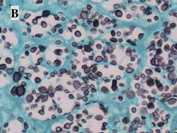
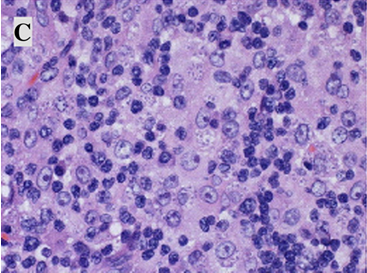
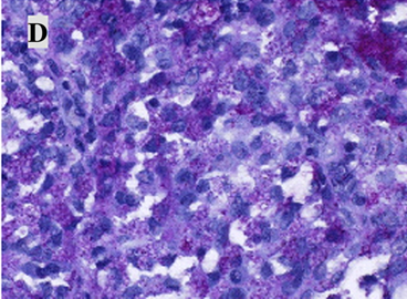
A 31-YOM had fever, abdominal pain and gingival lesion.


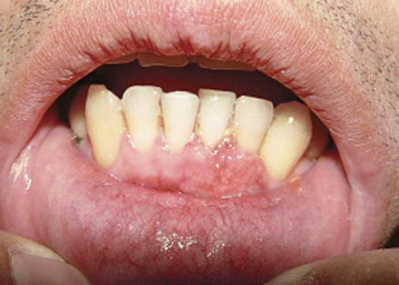
   ชายอายุ 31 ปีมีไข้และปวดทั่วท้องมา 5 วัน. ผู้ป่วยมีรอยโรคที่เหงือกและบวมๆ ที่คอซึ่งไม่เจ็บมา 1 เดือน อ่อนเพลียและเบื่ออาหารมา 3 เดือน. น้ำหนักลดไป 10 กก. ผู้ป่วยทำงานก่อสร้าง.
Physical examination revealed oral candidiasis, granulomatous gingiva (รูป 1), multiple palpable left supraclavicular and cervical lymph nodes (the largest up to 3.0 × 2.5 cm2, and hepatosplenomegaly.
Laboratory investigation revealed
-CBC: WBC 5,800 cell/cu mmL with 91% neutrophils and 4% lymphocytes, hemoglobin level of 11.2 g/dL, a platelet count of 180,000 cells/cu mm

1. การวินิจฉัยที่น่าจะเป็นคืออะไร
2. จะ manage อย่างไร


Posted by : cpantip , E-mail : (chpantip@medicine.psu.ac.th) ,
Date : 2012-02-17 , Time : 15:45:43 , From IP : 172.29.3.68


ความคิดเห็นที่ : 1


   1.Where is the localizing for the lesion?
From the history reveals 1.Abdominal pain 2.Gingival granuloma
From the PE reveal 1.oral candidiasis 2.granulomatous gingiva 3.multiple palpable left supraclavicular and cervical lymph nodes 4. hepatosplenomegaly.
From the CBC shows Lymphopenia
IMP : From both history and physical examination can not localizing for primary site of the disease.So the most commom should be the diffuse process or hematologic spreading.And the CBC shows lymphopenia that makes immunodeficiency host is suspected now!!!!
2.What is the cause?
Most likely in this case should be chronic infection that causes the diffuse process or hematologic spreading.But the diffuse process is more likely due to the hematologic spread should have high grade fever +/- chill
So it is not usually that,the normal immumne status man have chronic diffuse process like in this case.Adult man with low immune status host HIV must be the first suspection.
3.Organism?
for this patient the chronic infectious process may cause by.....
1.Bacteria : Mycoplasma spp. Actinomyces, Nocardia.......
2.Fungus : Candida,Histoplasma,Penicillium
But from the etiology in the patient with chronic diffuse infectious process in bacteria : Mycoplasma is most likely and in fungus : Histoplasma and Penicillium are most likely........It need the tissue and bone marrow biopsy to show the organism.
LAB IN NEED:
1.HIV screening test
2.tissue(lesion site) and bone marrow biopsy to show the organism.


Posted by : kenny , E-mail : (streptokinase@hotmail.com) ,
Date : 2012-02-21 , Time : 13:43:50 , From IP : 172.29.18.164


ความคิดเห็นที่ : 2


   ชายอายุ 31 ปีมีปัญหาดังต่อไปนี้
1, Fever and generalized abdominal pain 5 days.
2. Granulomatous gingiva 1 mo.
3. Multiple lymphadenopathy with hepatosplenomegaly.
5. Anorexia 3 mo with wt loss 10 kg.
6. Oral candidiasis,
7. Lymphopenia Absolute lymphocyte count 230 cell/cu mm

ปัญหา 5-7 เข้าได้กับ AIDS มากที่สุด. ผู้ป่วยรายนี้ CD4 ต่ำมาก (ถ้าคิด % ของ CD4 เป็น 10% ผู้ป่วยก็จะมี CD4 23 ตัว/cu mm เท่านั้น) จึงมีโอกาสเกิด opportunistic infection ต่างๆ ได้ง่าย โรคที่มีอาการและอาการแสดงดังในผู้ป่วยนี้ได้แก่ TB, MAC infection, systemic fungal infection (histoplasmosis, penicillosis marneffei, cryptococcosis). นอกจากนี้ ผู้ป่วยยังมีโอกาสเป็น malignant disease เช่น lymphoma ได้
Investigation ที่ควรทำคือ
1. HIV antibody
2. CXR
3. Liver function test + renal function test
และเพื่อหาเชื้อก่อโรค คือ
4. Lymph node biopsy
5. Gingival biopsy
6. Hemoculture for mycobacterium and fungi


Posted by : lara , Date : 2012-02-21 , Time : 14:23:59 , From IP : 172.29.27.98

ความคิดเห็นที่ : 3


   ขอโทษครับ mycobacterium ไม่ใช่ mycoplasma มึนหัวหมด

Posted by : Kenny , E-mail : (Streptokinase@hotmail.com) ,
Date : 2012-02-21 , Time : 16:39:19 , From IP : 110.49.232.87


ความคิดเห็นที่ : 4


   ขออนุญาติตอบเป็นภาษาไทยอีกครั้งนะครับ คอมเครื่องที่ใช้ตอนแรกมันเปลี่ยนภาษาไม่ได้ครับ
(1).ตำแหน่งของพยาธิสภาพที่เป็นไปได้มากที่สุดในผู้ป่วยรายนี้
จากการซักประวัติเราพบว่า 1.Abdominal pain 2.Gingival granuloma และจากการตรวจร่างกายพบว่า 1.oral candidiasis 2.granulomatous gingiva 3.multiple palpable left supraclavicular and cervical lymph nodes 4. hepatosplenomegaly. สังเกตได้ว่าจากประวัติและการตรวจร่างกายนี้ไม่สามารถบอกตำแหน่งของพยาธิสภาพได้ว่าอยู่ที่ตำแหน่งใดเพราะมีรอยโรคกระจัดกระจากไปทั่วร่างกายซึ่งส่วนมากจะเป็น solid organ เสียส่วนใหญ่ในที่นี้พยาธิสภาพอาจจะเป็นแบบกระจัดกระจายหรือในกระแสเลือดก็ได้ แต่ในผู้ป่วยรายนี้ผมคิดถึงพยาธิสภาพแบบกระจัดกระจายมากกว่าครับเพราะถ้าเป็นพยาธิสภาพที่ไปตามกระแสเลือดน่าจะมีความรู้แรงมากกว่านี้และเป็นในหลายๆอวัยวะมากกว่านี้ครับ
(2).อะไรทำให้เกิดพยาธิสภาพในผู็ป่วยรายนี้ : ในคนปรกติพยาธิสภาพแบบกระจัดกระจายนี้เกิดได้ยากครับการที่จะเกิดลักษณะอย่างนี้คงจะต้องมีปัจจัยส่งเสริมให้เกิดในชายหนุ่มสาเหตุที่จะต้องคิดถึงคือ immunodeficiency host ครับ โดยเมื่อเราดูประกอบกับระดับ Lymphocyte count ใน CBC ของผู้ป่วยรายนี้แล้วเราจะพบ Lymphopenia ซึ่งช่วยสนับสนุน immunodeficiency host ครับ โดยสาเหตุที่ทำให้เกิดในรายนี้ผมคิดถึงสาเหตุจาก infection มากที่สุดครับ
(3).Organism ที่ทำให้เกิด disseminated infection ที่ run แบบ chronic process ใน
Immunodeficiency host ได้แก่ Bacteria : Mycobacterium tuberculosis,Non-tuberculous mycobacterium,Actinomyces,Nocardia....... ส่วน Fungus : Candida,Histoplasma,Penicillium ครับ จะทราบชนิดของเชื้อได้คงต้องทำการเก็บตัวอย่าง
LAB
1.HIV screening test
2.Tissue(lesion site) and bone marrow biopsy to show the organism.
3.CXR + Liver function


Posted by : kenny , E-mail : (streptokinase@hotmail.com) ,
Date : 2012-02-21 , Time : 22:27:18 , From IP : 110.77.216.119


ความคิดเห็นที่ : 5


   ขอบคุณคุณหมอ Kenny และคุณหมอ Lara ค่ะ ที่ตอบมาถูกต้องแล้วค่ะ

-C-reactive protein level 49.2 mg/L,
-lactate dehydrogenase level of 211 U/L.
-Computed tomographic scan of the abdomen and chest was remarkable for diffused lymphadenopathy in the cervical, supraclavicular, mediastinal, para-aortic, paratracheal, mesenteric, and retroperitoneal space.


Posted by : cpantip , Date : 2012-02-23 , Time : 10:03:35 , From IP : 172.29.3.68

ความคิดเห็นที่ : 6


   Biopsy specimens from different body sites:
1. Bone marrow aspiration smear รูป B: Liu stain, × 1000


Posted by : cpantip , Date : 2012-02-23 , Time : 10:07:13 , From IP : 172.29.3.68

ความคิดเห็นที่ : 7


   2. Gingival biopsy, Giemsa stain, × 1000

Posted by : cpantip , Date : 2012-02-23 , Time : 10:08:40 , From IP : 172.29.3.68

ความคิดเห็นที่ : 8


   3. Cervical lymph nodes (C) hematoxylin-eosin stain, × 400

Posted by : cpantip , Date : 2012-02-23 , Time : 10:09:30 , From IP : 172.29.3.68

ความคิดเห็นที่ : 9


   4. nasopharynx (D) periodic acid-Schiff stain, × 400.

Posted by : cpantip , Date : 2012-02-23 , Time : 10:10:11 , From IP : 172.29.3.68

ความคิดเห็นที่ : 10


   Lactophenol cotton blue stain of the microbe isolated from Gingival biopsy (×400).

Posted by : cpantip , Date : 2012-02-23 , Time : 10:11:47 , From IP : 172.29.3.68

ความคิดเห็นที่ : 11


   A, Liu stain of bone marrow aspiration shows multiple oval yeasts with vacuolated cytoplasm (arrowhead; 1000×).
B, Giemsa stain of gingival biopsy specimen shows multiple oval yeasts with budding cells (arrowheads; 1000×).
C, Cervical lymph node, hematoxylin and eosin stain (400×).
D, Nasopharynx, periodic acid-Schiff stain (400×).

Lactophenol cotton blue stain of the mold showed several thick-walled, tuberculate macroconidia (arrowheads; 400×).


Posted by : cpantip , Date : 2012-02-23 , Time : 10:14:05 , From IP : 172.29.3.68

ความคิดเห็นที่ : 12


   การวินิจฉัย: Acquired immunodeficiency syndrome with disseminated histoplasmosis
การติดเชื้อ Histoplasma capsulatum สามารถทำให้เกิดอาการโรคได้หลายรูปแบบ และเป็นกับหลาย organ system โดยเชื้อไปทาง lymphatics และ bloodstream, เช่นเดียวกับ tuberculosis. Clinical presentations ของผู้ป่วย AIDS ที่มี disseminated histoplasmosis คือ fever, respiratory symptoms, weight loss, diarrhea, malaise, anorexia, hepatosplenomegaly และ lymphadenopathy. Oral involvement พบได้ไม่บ่อย แต่เมื่อพบ mucocutaneous lesion (รูป 1) ก็ต้องคิดถึงว่าเป็นอาการโรคของ systemic infection.
Disseminated histoplasmosis มีอาการโรคเหมือน disseminated mycobacterial infections และ lymphoma จึงมีโอกาสวินิจฉัยผิดได้ง่ายและทำให้ผู้ป่วยได้รับยาต้านเชื้อราช้า. ผู้ป่วยรายนี้ได้รับการวินิจฉัยเบื้องต้นว่าเป็น lymphoma เพราะมี diffused lymphadenopathy.


Posted by : cpantip , Date : 2012-02-23 , Time : 10:16:39 , From IP : 172.29.3.68

ความคิดเห็นที่ : 13


   Liu stain ของ bone marrow biopsy specimen พบ oval yeasts with obvious vacuolated cytoplasm (รูป 2). biopsy specimens พบ numerous intracytoplasmic และ extracellular oval yeasts, ขนาด 2–4 μm, มี budding (รูป 2-5). histological findings เช่นนี้พบได้ในการติดเชื้อที่เกิดจาก Histoplasma capsulatum, Blastomyces dermatidis, Candida glabrata, Cryptococcus neoformans, Penicillium marneffei, หรือ Leishmania species. การเพาะเชื้อราของ gingival biopsy specimens ขึ้น H. capsulatum (รูป 6). แม้ว่า Sepedonium species ก็มี tuberculate macroconidia เหมือนกัน, แต่แยกจาก H. capsulatum ได้เนื่องจาก Sepedonium species ไม่เป็น dimorphic และไม่ form microconidia.

Posted by : cpantip , Date : 2012-02-23 , Time : 10:17:24 , From IP : 172.29.3.68

ความคิดเห็นที่ : 14


   ผู้ป่วย disseminated histoplasmosis มักเสียชีวิตถ้าไม่ได้รับการรักษาด้วยยาต้านเชื้อราที่เหมาะสม. การรักษาเริ่มด้วย amphotericin B ขนาด 0.7–1.0 มก/กก หรือ lipid formulation ของ amphotericin B ขนาด 3–5 มก/กก แล้วตามด้วย oral itraconazole (200 มก. วันละ 2 ครั้ง) อย่างน้อย 12 เดือน. ในผู้ป่วยที่ได้รับ antiretroviral therapy ต้องระวัง interactions ระหว่าง itraconazole และ antiviral agents. ผู้ป่วยรายนี้ได้รับ zidovudine-lamivudine และ atazanavir ร่วมกับ oral itraconazole. หลังเริ่มการรักษา ต่อมน้ำเหลืองและรอยโรคที่เหงือกก็ยุบลง.

Ref. Huang C-C, et al. Clin Infect Dis. (2011) 53 (11): 1118-9, 1164-5.


Posted by : cpantip , Date : 2012-02-23 , Time : 10:19:49 , From IP : 172.29.3.68

สงวนสิทธิ์การแสดงความคิดเห็นสำหรับ สมาชิกเท่านั้น