ความคิดเห็นทั้งหมด : 6

A 47-YOM smoker presented to ER with chest pain and shortness of breath.


   ชายอายุ 46 ปีได้ไปตรวจที่ห้องฉุกเฉินเนื่องจากเจ็บหน้าอกและหายใจเหนื่อย. ผู้ป่วยสูบบุหรี่

PE: He was in mild distress with an oxygen saturation of 97% while breathing ambient air. Chest examination revealed hyperresonance and decreased breath sounds on the left.

1. การวินิจฉัยน่าจะเป็นอะไร
2. จะ manage อย่างไร


Posted by : cpantip , E-mail : (chpantip@medicine.psu.ac.th) ,
Date : 2011-12-01 , Time : 15:33:00 , From IP : pcache.psu.ac.th


ความคิดเห็นที่ : 1


   1) Pertinent data
1. sudden onset of chest pain and dyspnea
2. สูบบุหรี่
3. hyperresonance and decreased breath sounds on the left lung.

คิดถึง Left pneumothorax มากที่สุดค่ะ

2) Management
-CXR ด่วนคะ ถ้ามี pneumothorax จะได้ดูดลมออก หรือใส่ chest drain


Posted by : daisy1 , Date : 2011-12-04 , Time : 13:57:23 , From IP : 172.29.3.14

ความคิดเห็นที่ : 2


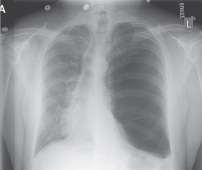
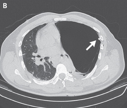
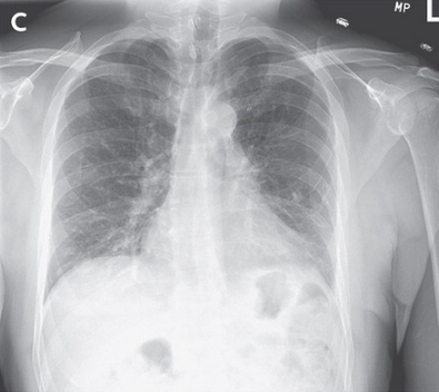
   คุณหมอ Daisy1 ตอบถูกแล้วค่ะ

แพทย์ได้ส่ง CXR (รูป A)

1. พบความผิดปกติอะไร
2. จะ manage อย่างไร


Posted by : cpantip , Date : 2011-12-08 , Time : 11:59:45 , From IP : 172.29.3.14

ความคิดเห็นที่ : 3


   Radiography of the chest showed a large lucency occupying the entire left hemithorax with contralateral mediastinal shift and left lung compressive atelectasis (รูป A).

ได้ใส่ chest tube แต่ follow-up radiography พบว่าปอดซ้ายไม่ขยาย.

จะ manage อย่างไรต่อไปคะ


Posted by : cpantip , Date : 2011-12-12 , Time : 13:49:20 , From IP : 172.29.1.48

ความคิดเห็นที่ : 4


   ใส่ chest tube แล้วปอดไม่ขยาย คิดถึง
1. มี bronchpleural fistula ควรมีลมออกมาในขวดปุดๆๆ
2. เป็น lung bullus ขนาดใหญ่

คิดว่าน่าจะทำ CT scan of lung ค่ะ


Posted by : daisy1 , Date : 2011-12-14 , Time : 12:04:04 , From IP : 172.29.1.48

ความคิดเห็นที่ : 5


   ได้ส่ง Computed tomography (รูป B)
Computed tomography showed a giant tension bulla occupying the entire left hemithorax and the chest tube in place within the left pleural space, external to the bulla.


Posted by : cpantip , Date : 2011-12-15 , Time : 10:24:37 , From IP : 172.29.1.48

ความคิดเห็นที่ : 6


   ผู้ป่วยได้รับการผ่าตัด bullectomy ซึ่งได้ผลดี มี reexpansion ของปอดซ้าย (รูป C).
การแยกระหว่าง pneumothorax และ giant bulla ทำได้ยาก. ถ้าเป็น pneumothorax, ปอดจะยุบไปสู่ hilum ข้างเดียวกัน ยกเว้นว่ามี adhesions ทำให้ปอดเคลื่อนไม่ได้. chest tube ที่ใส่เข้าไปใน pleural space ขับลมออกไปอย่างรวดเร็วทำให้ปอดขยายออก. ถ้าเป็น tension bulla, เนื้อปอดจะถูกกดให้อยู่รอบ bulla และ chest tube ถูกจากภายนอกก็จะอยู่นอก bulla, ดังที่พบในผู้ป่วยรายนี้.

Reference: Vega MA, et al. N Engl J Med 2011; 365:1915


Posted by : cpantip , Date : 2011-12-15 , Time : 10:26:22 , From IP : 172.29.1.48

สงวนสิทธิ์การแสดงความคิดเห็นสำหรับ สมาชิกเท่านั้น