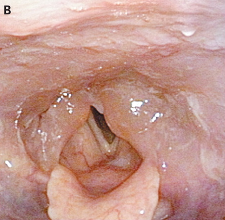
Fiberoptic laryngoscopy: marked mucosal edema of the arytenoid region (รูป A, black arrows), aryepiglottic fold (arrowhead), and posterior wall of the pharynx (white arrows).
ผู้ป่วยได้รับ IV hydrocortisone.
หนึ่งชั่วโมงต่อมา laryngopharyngeal edema ยุบลงบางส่วน. (รูป B).
วันรุ่งขึ้น ผู้ป่วยไม่มีอาการผิดปกติใดๆ fiberoptic laryngoscopy พบว่ามี minor edema ของ left arytenoid mucosa.
Posted by : cpantip , Date : 2011-11-20 , Time : 13:59:16 , From IP : 172.29.3.68
|