A 53 YOM had tender RUQ abdominal mass for 4 wksA 53-year-old man presented with a four-week history of abdominal pain and a tender, nonerythematous mass (5 by 5 cm) in the right upper quadrant. He had a temperature of 36.3°C, a leukocyte count of 8100 per cubic millimeter, and normal liver-function tests. What is/are abnormalities found in his CT scan? Posted by : chpantip , E-mail : (chpantip@medicine.psu.ac.th) , Date : 2008-06-19 , Time : 08:26:40 , From IP : 172.29.3.68 |
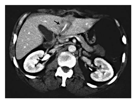
Finding - Hypoechoic lesion at segment 3 with calcified lesion and perihepatic inflammation (anterior) Posted by : Nongnham , E-mail : (Nongnham025@gmail.com) , Date : 2008-06-20 , Time : 06:51:33 , From IP : 222.123.139.86 |
CT finding : hypodense lesion , well define border , half of Lt lobe of liver with abnormal hyperdense(bony level ) pin shape inside , and also lesion extensed to skin Imaging diagnosis : Liver abscess with inside FB Clinical diagnosis 1.External trauma : น่าจะได้ประวัติ 2.Parasistic visceral larva migran : ไม่น่าจะมี hyperdense pin-shape lesion ( มี hyperdense ได้แต่ไม่น่าจะhyperdense ถึง bony level ) 3.Bias with reading experience : fishbone from raptured 1st part of duodenum with liver abscess Treatment Drainage due to 1.Lt lobe : small , near vital organ 2.for definite diagnosis 3.Remove FB ATB ถ้าเป็น fish bone จริงให้ ATB cover enterobacteriacae ( GNB and enterococci ) : Gentamicin +Ampicillin Posted by : เชน , E-mail : (sarunyouchusri@hotmail.com) , Date : 2008-06-20 , Time : 12:09:06 , From IP : 172.29.3.68 |
ขอบคุณคุณหมอเชน และ คุณหมอ Nongnham มากค่ะ ขอเล่าความก้าวหน้าและขอถามต่อค่ะ CT scan of abdomen showed a mass in the left lobe of the liver with a thin, linear calcification and invasion beyond the capsule to the abdominal wall. The linear density within the abscess (arrow) was initially thought to be an artifact. ทำ CT-guided biopsy ได้หนอง 14 มล. เอาหนองมาย้อม Gram stain พบ gram-positive cocci in clusters, pairs, and chains and gram-positive and gram-negative rods. 1. ท่านคิดว่าเป็นเชื้ออะไร และจะให้ยาปฏิชีวนะใดแก่ผู้ป่วย Gentamicin +Ampicillin เพียงพอหรือไม่ 2. ต้อง drain หนองหรือไม่ percutaneous drainage เพียงพอไหม Posted by : chpantip , E-mail : (chpantip@medicine.psu.ac.th) , Date : 2008-06-26 , Time : 11:02:26 , From IP : 172.29.3.68 |
ขอโทษค่ะที่ไม่ได้เขียน progression ของผู้ป่วยรายนี้ เพิ่งมาเปิดพบ ขอตอบคำถามนะคะ Gram stain ของหนอง พบ gram-positive cocci in clusters, pairs, and chains and gram-positive and gram-negative rods. 1. ท่านคิดว่าเป็นเชื้ออะไร และจะให้ยาปฏิชีวนะใดแก่ผู้ป่วย Gentamicin +Ampicillin เพียงพอหรือไม่ ตอบ ลักษณะของหนองเป็น mixed infection ซึ่งต้องนึกถึง mixed aerobic-anaerobic infection ดังนั้น ถ้าให้ Gentamicin+ampicillin dHต้องให้ยาที่ ครอบคลุม Bactroides fragilis คือ metronidazole หรือ clindamycin ด้วย แต่ metronidazole ราคาถูกกว่าและมีประสิทธิภาพสูงกว่า 2. ต้อง drain หนองหรือไม่ percutaneous drainage เพียงพอไหม ผู้ป่วยรายนี้ได้รับการรักษาด้วย intravenous vancomycin and piperacillintazobactam (vancomycin สำหรับ resistant Gram-positive cocci เช่น MRSA และ resistant enterococci ถ้าเป็นผู้ป่วยไทย ให้ pip/tazo ก็พอ) เนื่องจากอาการของผู้ป่วยไม่ดีขึ้น จึงทำ surgical drainage พบว่ามีก้างปลาชิ้นบางๆอยูในฝี (ก้างปลาทะลุ duodenum ส่วนแรกไปเข้าตับกลีบซ้ายทางด้านหลัง) ผลการเพาะเชื้อจากหนองขึ้น Eikenella corrodens (เชื้อนี้พบได้ในปากของคน) ผู้ป่วยมีอาการดีขึ้นอย่างรวดเร็วหลังเอาก้างปลาออก ผู้ป่วยกลับบ้านโดยได้รับยา amoxicillinclavulanate รูปกิน เขาจำไม่ได้ว่าได้กลืนก้างปลาไปเมื่อใด เรื่องและรูป ได้มาจาก Kessler A.T., Kourtis A.P. Liver Abscess Due to Eikenella corrodens from a Fishbone. N Engl J Med 2001;345:e5. Posted by : chpantip , E-mail : (chpantip@medicine.psu.ac.th) , Date : 2008-07-17 , Time : 14:13:28 , From IP : 172.29.3.68 |