ความคิดเห็นทั้งหมด : 5

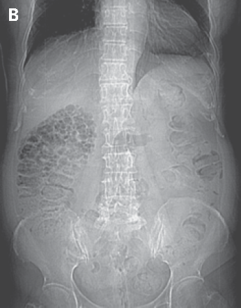
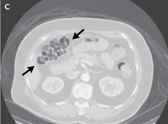
A 67-YOW had multiple polypoid lesions in the wall of ascending colon.


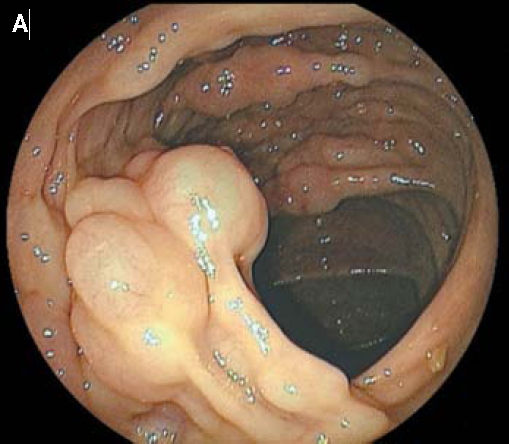
   หญิงอายุ 67 ปีซึ่งไม่มีอาการใดๆ ได้รับการทำ screening colonoscopy ซึ่งพบ multiple translucent polypoid lesions ในผนังของ ascending colon (รูป A ).

1. การวินิจฉัยคืออะไร
2. จะ manage อย่างไร


Posted by : cpantip , E-mail : (chpantip@medicine.psu.ac.th) ,
Date : 2011-09-05 , Time : 12:10:42 , From IP : 172.29.3.68


ความคิดเห็นที่ : 1


   1.Dx : Pneumatosis Intestinalis because of translucent appearance.
Polypoid lesions ใน colon ก็ควรจะนึงถึง CA colon เป็นอันดับแรกเพราะเป็นโรคที่พบได้บ่อยและมีความรุนแรงถึงชีวิต รองลงมาก็อาจจะเป็น Carcinoid,Neuroendocrine Tumors,Gastrointestinal Lymphoma โดยทั้งหมดนี้สามารถให้การวินิจฉัยจาก colonoscope ได้ แต่โดยปรกติแล้ว tumor ของ colon มักจะเป็น SOLID mass ยกตัวอย่างที่พบมากที่สุดคือ Colon adenocarcinoma ซึ่งมี 3 รูปแบบ ได้แก่ Sessile Ulcer และ Polyp ซึ่งล้วนเป็น SOLID mass แต่คนไข้คนนี้เป็นลักษณะของ translucent สิ่งที่อยู่ภายในควรจะเป็น ลม หรือ น้ำ แต่โดยปรกติที่พบทั่วไปจะเป็นสมเสียมากกว่า แต่โดยทั่วไปของ Pneumatosis Intestinalis ที่พบมักเกิดจาก Venous plexus ใน Bowel เกิด congestion ทำให้เกิด Bowel layer Ischemia และ Necrosis ทำให้ลมใน Bowel เข้าไป fill ในชั้นนั้นเกิดเป็น Pneumatosis Intestinalis ผู้ป่วยจริงควรมีอาการบ้าง ผมจึงไม่ค่อยแน่ใจเท่าไรครับ แต่ขอ Dx ตาม Key word ที่ว่า TRANSLUCENT ครับ
2.ถ้าเป็นไปได้อยาก Biopsy เลยแต่ไม่ทราบว่าจะทำได้ไหม ทำถุงลมแตกจะเป็นอะไรไหม ถ้า Biopsy ไม่ได้ก็อยากส่งเป็น BioMarker เช่น CEA +/- CA19-9 ดูครับ แต่จากความคิดเล้วถ้า Biopsy ได้จะดีมากครับเพราะยังไงก็แหย่ Colono ไปแล้ว


Posted by : kenny , E-mail : (Streptokinase@hotmail.com) ,
Date : 2011-09-07 , Time : 02:17:28 , From IP : 110.77.230.62


ความคิดเห็นที่ : 2


   คุณหมอ Kenny ตอบได้ดีค่ะ และให้การวินิจฉัยถูกต้อง

รายนี้ แพทย์ส่ง Computed tomographic imaging of the abdomen

CT scan of abdomen revealed a honeycomb-like cluster of air-filled sacs within the right colonic wall (รูป B, scout image; และรูป C, axial image, arrows), เข้าได้กับ pneumatosis cystoides intestinalis.


Posted by : cpantip , Date : 2011-09-08 , Time : 13:35:37 , From IP : 172.29.3.68

ความคิดเห็นที่ : 3


   การมี air-filled blebs อยู่ในชั้น subserosal และ submucosal อาจเป็น benign finding โดยไม่มีสาเหตุที่ทำให้เกิดที่ชัดเจน. ในทางตรงกันข้าม ภาวะนี้อาจเป็นอาการแสดงของโรคที่รุนแรงหลายอย่างซึ้งได้แก่ gastrointestinal infection, inflammatory bowel disease, life-threatening intraabdominal catastrophe (เช่น bowel infarction หรือมี underlying malignant condition), organ transplantation, และ obstructive lung disease.

ในผู้ป่วยรายนี้ การเกิด pneumatosis cystoides intestinalis สัมพันธ์กับการใช้ยาชนิดหนึ่ง ยานั้นคืออะไรคะ


Posted by : cpantip , Date : 2011-09-08 , Time : 13:37:10 , From IP : 172.29.3.68

ความคิดเห็นที่ : 4


   ลืมบอกไปว่า ผู้ป่วยเป็นเบาหวาน type 2 ด้วยค่ะ

Posted by : cpantip , Date : 2011-09-09 , Time : 15:59:50 , From IP : 172.29.3.68

ความคิดเห็นที่ : 5


   มี case reports หลายรายที่มี pneumatosis cystoides intestinalis สัมพันธ์กับการใช้ alpha-glucosidase inhibitors. มีผู้ตั้งสมมุติฐานว่า แก๊ซที่ถูกสร้างขึ้นจากการ ferment carbohydrate ที่มีมากมายใน lumen ของลำไส้ ได้เข้าไปสู่เนื้อเยื่อของผนังลำไส้. ผู้ป่วยรายนี้ได้รับ alpha-glucosidase inhibitor acarbose ด้วยสำหรับการรักษา type 2 diabetes mellitus. แพทย์ได้แนะนำให้ผู้ป่วยหยุดยา acarbose.

สามเดือนต่อมา การทำ plain radiograph of the abdomen เพื่อติดตาม พบว่า intramural air หายไปหมด.

Reference: Wu S-S, et al. Pneumatosis cystoides intestinalis.
N Engl J Med 2011; 365:e16.


Posted by : cpantip , Date : 2011-10-06 , Time : 11:44:36 , From IP : 172.29.3.14

สงวนสิทธิ์การแสดงความคิดเห็นสำหรับ สมาชิกเท่านั้น