ความคิดเห็นทั้งหมด : 7

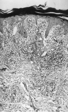
A 21-YOF alcoholic had fever and painful ulceronecrotic skin lesions for 3 wks.


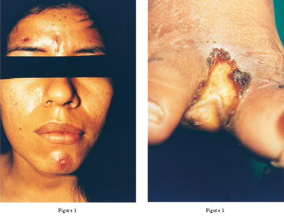
   หญิงอายุ 21 ปี ติดสุรา มารพ.เนื่องจากมีไข้และรอยโรคที่ผิวหนังมา 3 เดือน . ผู้ป่วยไม่ใช้ยาเสพติดใดๆ แต่ให้ประวัติว่ามีเพศสัมพันธ์โดยไม่ได้ป้องกันกับคู่นอนหลายคน. ผู้ป่วยสบายดีจนเมื่อ 3 สัปดาห์ก่อน ก็เริ่มมีไข้ และมีรอยโรคที่ผิวหนังซึ่งเป็น multiple painful ulceronecrotic skin lesions ที่แขนขาและที่หน้า (รูป 1 และ 2). ไม่มีรอยโรคที่ฝ่ามือฝ่าเท้า.
การย้อม discharge จากแผล ด้วย Gram stain, Wright stain และ acid fast stain ไม่พบเชื้อใดๆ.
1. การวินิจฉัยคืออะไร
2. จะ manage อย่างไรต่อไป


Posted by : cpantip , E-mail : (chpantip@medicine.psu.ac.th) ,
Date : 2011-07-22 , Time : 10:10:18 , From IP : 172.29.3.68


ความคิดเห็นที่ : 1


   ขอโทษค่ะ ผู้ป่วยมีไข้และรอยโรคที่ผิวหนังมา 3 สัปดาห์ค่ะ

Posted by : cpantip , Date : 2011-07-22 , Time : 10:12:00 , From IP : 172.29.3.68

ความคิดเห็นที่ : 2


   Primary syphilis kub

Posted by : kenny , E-mail : (streptokinase@hotmail.com) ,
Date : 2011-07-22 , Time : 12:30:53 , From IP : 172.29.27.95


ความคิดเห็นที่ : 3


   ต้องขอโทษอาจารย์ด้วยที่ตอนแรกตอบมาสั้นๆเพราะตอบมาทางโทรศัพท์และพิมพ์ภาษาไทยไม่ได้ครับ
CC:fever and painful ulceronecrotic skin lesions for 3 wks.
S:
1. หญิงอายุ 21 ปี ติดสุราและมีเพศสัมพันธ์โดยไม่ได้ป้องกันกับคู่นอนหลายคน
2. ไข้และรอยโรคที่ผิวหนังมา 3 เดือน
O:
1. multiple painful ulceronecrotic lesion ที่แขนขาและที่หน้าไม่มีรอยโรคที่ฝ่ามือฝ่าเท้า
2. dischargeจากแผลด้วยGram stain,Wright stainและacid fast stain ไม่พบเชื้อใดๆ
A: ในคนนี้ผมคิดถึง Primary syphilis มากที่สุดเพราะว่าคนไข้มีไข้และมีรอยโรคในทางระบบผิวหนังทำให้คิดถึงการติดเชื้อโดยที่การติดเชื้อนั้นมีผลทั่วทั้งร่างกายครับ และแน่นนอนว่าการติดเชื้อที่ผิวหนังย่อมเกิดจาก Bacteria Virus Fungus or Parasite ในที่นี้ผมคิดถึง bacteria มากที่สุดเพราะว่าลักษณะรอยโรคทางผิดหนังที่เกิดจาก parasite Fungus หรือ Dermatophyte นั้นไม่เหมือนเลยครับยิ่ง dermatophyte นั้นจะอยู่แค่ในชั้นที่เป็นKeratinจะไม่invadeครับ แต่ถ้าเป็นไวรัสอาการนั้นมันนานเกินไปครับและ skin viral infection ที่ติดแล้วมีไข้เด่นก็มีไม่กี่ตัวและรอยโรคที่ผิวหนังก็ไม่เหมือนด้วยครับ Bacteria เป็นตัวที่คิดถึงมากที่สุด จากระยะเวลาการเกิดที่นานทำให้ผมคิดถึงกลุ่มของ Spirochete มากที่สุดและเมื่อประกอบกับประวัติเรื่องเพศสัมพันธ์แล้วทำให้คิดถึงมากที่สุด Treponema pallidum เป็นตัวที่ผมคิดถึงครับ โดยเมื่อดูจากแผลที่ผิวหนังแล้ว multiple painful ulceronecrotic lesion ก็ตรงไปตรงมาว่าน่าเป็น Primary syphilis แต่โดยปรกติแล้ว primary Syphilis มักจะมีแผลเป็นลักษณะที่เดียว single firm, painless, non-itchy skin ulceration with a clean base and sharp borders แต่เท่าที่รู็นั้นทราบว่าร้อยละ40นั้นสามารถเกิดเป็น multiple lesion ได้และมักจะสัมพันธ์กับการติดHIVครับ
P:
1.ผมคิดว่าควรจะต้องแก้ไขปัญหาที่ต้นเหตุก่อนครับในที่นี้ก็คือ เพศสัมพันธ์ที่เหมาะสมและถูกต้องปลอดภัยผมคิดว่าเราควรสอนผู้ป่วย เพื่อนให้ปลอดภัยกับตัวผู้ป่วยเองและงดการแพร่เชื้อต่อไป
2.คนนี้ควรทำHIV screening test เพราะว่า multiple lesion on primary syphilis นั้นมีความเกี่ยวข้องกัยการติด HIV สูง
3.ผมคิดว่าควรทำ urine preg test ครับเพื่อการรักษาระยะยาวและความปลอดภัย
4.Antibiotics The first-choice ควรเป็น PenicillinG IM ครับ
5.หลังจากที่เราเริ่มยาไปได้ 24 ชั่วโมงเราควรตามอาการคนไข้อย่างใกล้ชิดเพื่อเฝ้าระวัง
Jarisch-Herxheimer reaction ซึ่ีงเป็น immune system in response to lipoproteins จาก rupturing syphilis bacteria โดยผู้ป่วยมักจะมีอาการfever, muscles pains, headache, and tachycardia


Posted by : kenny , E-mail : (Streptokinase@hotmail.com) ,
Date : 2011-07-22 , Time : 19:40:04 , From IP : 119.42.83.191


ความคิดเห็นที่ : 4


   5.modified Steiner silver stain. discharge จากแผล ถ้าเป็นจริงอาจจะพบ spiral-shaped, Gram-negative, highly mobile bacterium

Posted by : kenny , E-mail : (Streptokinase@hotmail.com) ,
Date : 2011-07-22 , Time : 19:44:41 , From IP : 119.42.83.191


ความคิดเห็นที่ : 5


   คุณหมอ Kenny ตอบถูกแล้วค่ะ

Diagnosis: Malignant Syphilis
Serological tests for HIV infection ได้ผลบวก และ CD4 cell count 272/cumm (21%).
ผลการเพาะเชื้อ bacteria, fungi, และ mycobacterium ของ skin biopsy specimens were ได้ผลลบ.

ในผู้ป่วยรายนี้ histopathologic findings ของ skin biopsy specimen (รูป 3) ทำให้แพทย์นึกถึง syphilis. VDRL titer 1:512 และ positive microhemagglutination assay with Treponema pallidum ยืนยันการวินิจฉัย syphilis.

ผู้ป่วยตอบสนองดีต่อการรักษาด้วย IV penicillin G นาน 14 วัน skin lesions และไข้หายไปหมด. มี Jarisch-Herxheimer reaction ในวันแรกของการรักษา.


Posted by : cpantip , Date : 2011-07-25 , Time : 10:35:20 , From IP : 172.29.3.68

ความคิดเห็นที่ : 6


   Figure 3. Microscopic examination of a skin biopsy specimen from a female patient with malignant syphilis shows proliferation of capillaries, interstitial edema, and a mixed inflammatory cell infiltrate
(stain, hematoxylin-eosin; original magnification, 1125).


Posted by : cpantip , Date : 2011-07-25 , Time : 10:47:40 , From IP : 172.29.3.68

ความคิดเห็นที่ : 7


   term ‘‘malignant syphilis’’ หรือ ‘‘syphilis praecox’’ ถูกใช้เรียก clinical form ของ secondary syphilis ซึ่งมีลักษณะสำคัญคือ ไข้, malaise, และ ulcerating, pustular และ/หรือ crusted skin lesions. Histopathologic examination: perivascular plasma cell infiltrates, epithelioid and giant cell granulomas, and swelling and proliferation of endothelial cells. อย่างไรก็ตาม พบ treponemes น้อยเพราะมี inflammatory response รุนแรง และมี spirochetes อยู่น้อย.

Reference: Wappner D, et al. Clinical Infectious Diseases 1997;25:1343, 1447.


Posted by : cpantip , Date : 2011-07-25 , Time : 10:49:51 , From IP : 172.29.3.68

สงวนสิทธิ์การแสดงความคิดเห็นสำหรับ สมาชิกเท่านั้น