ความคิดเห็นทั้งหมด : 3

A 43-YOM had lethargy, weakness, and headaches for 2 years.


   ชายอายุ 43 ปีซึ่งเป็นฝาแฝดแบบ identical twin มารพ.เนื่องจากเซื่องซึม อ่อนแรง และปวดหัวมานาน 2 ปี.

จากรูปถ่ายของผู้ป่วยและฝาแฝดของเขา
1. คนใดเป็นผู้ป่วย
2. บอกได้จากลักษณะอะไรบ้าง


Posted by : cpantip , E-mail : (chpantip@medicine.psu.ac.th) ,
Date : 2010-07-01 , Time : 11:01:14 , From IP : 172.29.3.68


ความคิดเห็นที่ : 1


   คิดว่าคนด้านขวามือ (คนที่ห่างไกลจากตัว A) ครับ เพราะไม่มีขน ที่หน้าอก ไม่มีหนวด และมีไขมันมากไป ไม่ค่อยมีกล้ามเนื้อ
ผู้ป่วยน่าจะมี pituitary tumor ทำให้เกิดอาการปวดหัวเรื้อรังได้ ถ้าก้อนกดเบียดเนื้อ pituitary ที่ดี ทำให้ไม่สามารถหลั่ง Hormone ได้ปกติ เกิด Panhypopituitarism or จำเพาะบาง Hormoen 2 nd hypogonad ก็ได้ครับ
work up MRI brain เน้นดู pituitary gland


Posted by : nimbus , Date : 2010-07-05 , Time : 21:40:50 , From IP : 61.7.241.125

ความคิดเห็นที่ : 2


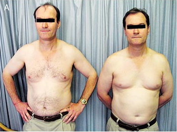
   คุณหมอ nimbus เก่งมากค่ะ ที่ตอบมาถูกต้องแล้วนะคะ ผู้ป่วยคือคนที่เตี้ยกว่าและยืนทางขวามือ

ผู้ป่วยรายนี้ยังมีอาการขนตามตัวค่อยๆ ร่วงและ ความต้องการทางเพศค่อยๆลดลงมานาน 2 ปี.
เมื่ออายุ 6 ปี ผู้ป่วยได้รับการผ่าตัด left orchidopexy.
Physical examination revealed central adiposity, nipple pallor, proximal muscle wasting, preservation of scalp hair, loss of body hair, gynecomastia, and right and left testicular volumes of 12 ml and 5 ml, respectively (normal volume, >15).

จากการตรวจร่างกายแสดงถึงลักษณะของ hypopituitarism และhypogonadism ของผู้ป่วย (คนยืนทางขวามือ) ซึ่งเห็นได้ชัดเจนเมื่อเทียบกับคู่แฝดของเขา (คนยืนทางซ้ายมือ).

Laboratory tests ของผู้ป่วยยืนยันว่าเป็น secondary hypogonadism:
1. testosterone level of 1.5 nmol per liter (ค่าปกติ, 9.9 to 27.8),
2. follicle-stimulating hormone level of 2.8 IU per liter (ค่าปกติ, 1.5 to 12.4), และ
3. luteinizing hormone level of 1.5 IU per liter (ค่าปกติ, 1.7 to 8.6).

ส่วน serum prolactin levels, thyroid function (วัด free thyroxine และ thyroid-stimulating hormone), และ cortisol levels อยู่ในเกณฑ์ปกติ; insulin-like growth factor I level ลดลงเล็กน้อย.


Posted by : cpantip , Date : 2010-07-07 , Time : 10:27:00 , From IP : 172.29.3.68

ความคิดเห็นที่ : 3


   Magnetic resonance imaging of the pituitary showed a lobulated, contrast-enhancing suprasellar mass (16 x 29 mm; seen in the coronal view in Panel B [arrow] and in the sagittal view in Panel C [arrow]).

หลังทำ needle biopsy และต่อมาก็ทำ excision โดย craniotomy, pathological analysis ยืนยันการวินิจฉัย pituicytoma.

หลังการผ่าตัด นอกเหนือไปจากชัก 1 ครั้ง ผู้ป่วยอาการดีขึ้นโดยต้องได้รับ pituitary-replacement therapy และอาการผิดปกติต่างๆ ค่อยๆหายไป

Reference: Newnham HH, Rivera-Woll LM. Hypogonadism due to pituicytoma in an identical twin. NEJM 2008;359 (26): 2824.


Posted by : cpantip , Date : 2010-07-07 , Time : 10:30:39 , From IP : 172.29.3.68

สงวนสิทธิ์การแสดงความคิดเห็นสำหรับ สมาชิกเท่านั้น