ความคิดเห็นทั้งหมด : 2

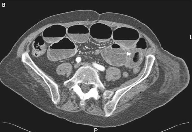
A 72-YOW had abdominal pain with nausea & vomiting for 2 days.


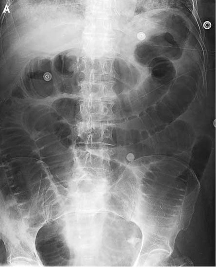
   หญิงอายุ 72 ปีถูกส่งตัวมารพ.เนื่องจากปวดท้องมานาน 2 วันร่วมกับคลื่นไส้และอาเจียน. ในเวลา 10 ปีที่ผ่านมา ผู้ป่วยมีโรค Alzheimer ซึ่งเป็นมากขึ้น ทำให้ผู้ป่วยต้องอยู่ใน long-term care facility.
On physical examination, there were no abdominal scars or umbilical, inguinal, or femoral hernias.
Laboratory tests revealed a normal white-cell count.

1. ในรูป A คือ abdominal radiologic examination พบความผิดปกติอะไรบ้าง
2. การวินิจฉัยที่น่าจะเป็นคืออะไร
3. จะ manage อย่างไรต่อไป


Posted by : cpantip , E-mail : (chpantip@medicine.psu.ac.th) ,
Date : 2010-06-06 , Time : 14:12:13 , From IP : 172.29.3.68


ความคิดเห็นที่ : 1


   1. ในรูป A คือ abdominal radiologic examination พบความผิดปกติคือ small bowel dilate มากทั่วๆ ไป
2. การวินิจฉัยที่น่าจะเป็นคือ small bowel obstruction ค่ะ
3. management
- ส่ง CT abdomen เพื่อหาว่ามีก้อนอุดตันที่ไหน
-ใส่ NG tube เพื่อ decompress bowel.
-ปรึกษาศัลยกรรม


Posted by : lara , Date : 2010-06-10 , Time : 15:16:01 , From IP : 172.29.3.68

ความคิดเห็นที่ : 2


   คุณหมอ lara ตอบถูกแล้วนะคะ

The abdominal radiologic examination was suggestive of a complete small-bowel obstruction (รูป A).

ผู้ป่วยได้รับการตรวจ CT abdomen
Computed tomography showed small-bowel obstruction by an intraluminal mass (รูป B, arrow). This mass had a hyperdense periphery and an aerated core.
ระหว่างการผ่าตัด laparotomy, แพทย์ได้ทำ enterotomy และพบลูกบอลพลาสติก อยูในรูลำไส้. ลูกบอลมีขนาดเส้นผ่าศูนย์กลาง 4 ซม.
จากการสอบถามผู้ป่วย ไม่ได้ข้อมูลว่าผู้ป่วยตั้งใจกินลูกบอลเอง หรือเกิดขึ้นตั้งแต่เมื่อไร. ผู้ป่วยหายดีเป็นปกติ.

Reference: Dedouit F, Otal P. Small-Bowel Obstruction. NEJM 2008;358 (13): 1381.


Posted by : cpantip , Date : 2010-06-11 , Time : 13:56:20 , From IP : 172.29.3.68

สงวนสิทธิ์การแสดงความคิดเห็นสำหรับ สมาชิกเท่านั้น