ความคิดเห็นทั้งหมด : 2

An 83-YOM had painful, progressive enlarging mass in Rt subcostal region for 3 mo.


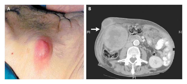
ชายอายุ 83 ปีมารพ.เนื่องจากมีก้อนที่ชายโครงขวาซึ่งเจ็บและโตขึ้นเรื่อยๆ มานาน 3 เดือน. ผู้ป่วยไม่มีอาการอื่นๆ และไม่มีประวัติการผ่าตัดใดๆ มาก่อน
Local examination revealed a tender, erythematous, fluctuant mass, 3 cm by 2 cm, with clinically significant surrounding induration and an underlying fixed mass (รูป A).

Computed tomography revealed that the mass was communicating with a large gall bladder mass (รูป B, arrow).

1. การวินิจฉัยที่น่าจะเป็นมากที่สุดคืออะไร
2. จะ manage อย่างไรต่อ


Posted by : cpantip , E-mail : (chpantip@medicine.psu.ac.th) ,
Date : 2010-04-29 , Time : 11:51:41 , From IP : 172.29.3.68


ความคิดเห็นที่ : 1


   ปัญหาของผู้ป่วยชายอายุ 83 ปี
1. painful, progressive enlarging mass in Rt subcostal region for 3 mo.
2. the mass was communicating with a large gall bladder mass from CT scan

ลักษณะของก้อน: tender, erythematous, fluctuant mass, 3 cm by 2 cm, with clinically significant surrounding induration เข้าได้กับ abscess ที่หน้าท้อง. ประวัติที่เป็นนาน 3 เดือนทำให้นึกถึง chronic infection ที่เกิดจาก M. tuberculosis, NTM, melioidosis, fungi, nocardia, actinomyces, etc.
ก้อนนี้ communicating with a large gall bladder mass ด้วย ก็ต้องคิดว่าการติดเชื้อน่าจะเริ่มต้นที่ถุงน้ำดีแล้วลุกลามมาเป็นฝีที่ผนังหน้าท้อง ที่ถุงน้ำดีอาจมีการอุดตันของ cystic duct จากนิ่วหรือ malignant tumor (เพราะผู้ป่วยสูงอายุมาก)
Management ควรทำ needle aspiration/biopsy ก้อนที่ผนังหน้าท้องและที่ gallbladder under ultrasound guide ถ้าได้หนองก็ย้อม Gram, modified acid fast และ acid fast และส่ง culture for aerobic bacteria และ for mycobacterium ก่อนให้การรักษา.


Posted by : lara , Date : 2010-05-01 , Time : 14:12:44 , From IP : 172.29.3.68

ความคิดเห็นที่ : 2


   คุณหมอ lara ตอบถูกแล้วค่ะ

Ultrasonography-guided needle biopsy, along with histologic and immunocytochemical examinations, confirmed that the mass was an adenocarcinoma of the gall bladder.

ผู้ป่วยไม่ต้องการ investigation หรือการรักษาใดๆต่อไป จึงกลับบ้านและได้รับการรักษาแบบ palliative care. ผู้ป่วยเสียชีวิต 5 สัปดาห์ต่อมา.

Reference: Rajagopalan S, Loudon M. Abdominal-Wall Abscess. NEJM 2010 ; 362 (8): e24.


Posted by : cpantip , Date : 2010-05-01 , Time : 16:16:09 , From IP : 172.29.3.68

สงวนสิทธิ์การแสดงความคิดเห็นสำหรับ สมาชิกเท่านั้น