
คุณหมอ lara ตอบถูกแล้วค่ะ ขอบคุณมากค่ะที่ติดตามและส่งคำตอบมาโดยตลอด
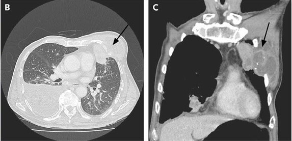
Computed tomography of the thorax showed a left-chest-wall collection of fluid with erosion of the ribs and extension into the pleural space, anterior mediastinum, and lung parenchyma (รูป B และ C, arrows). There were also bilateral nodular infiltrates, a right pleural effusion, and enlarged mediastinal lymph nodes.
ทำ fine-needle aspiration ที่ left chest wall ได้หนองออกมา ย้อมพบ acid-fast bacilli, และผลการเพาะเชื้อขึ้น Mycobacterium tuberculosis. การเพาะเชื้อของ bronchoalveolar-lavage fluid ก็ขึ้น M. tuberculosis และพบ granulomas ใน transbronchial biopsy.
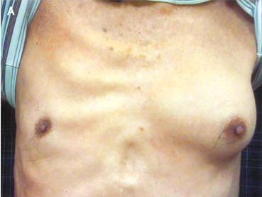
การวินิจฉัย คือ pulmonary tuberculosis with a tuberculous abscess of the left chest wall.
ผู้ป่วยได้รับการรักษาด้วยยารักษาวัณโรคและทำ débridement of the chest-wall abscess. หลังได้รับยารักษาวัณโรคนาน 9 เดือน ผู้ป่วยได้รับการทำ CT chest ซ้ำซึ่งผลพบว่ารอยโรคของวัณโรคหายไปหมด แลtผู้ป่วยสบายดี.
Reference: Koh J, Tee A. Tuberculous Abscess Manifesting as Unilateral Gynecomastia. NEJM 2009;361 (23): 2270.
Posted by : cpantip , Date : 2010-04-29 , Time : 13:38:56 , From IP : 172.29.3.68
|