ความคิดเห็นทั้งหมด : 6
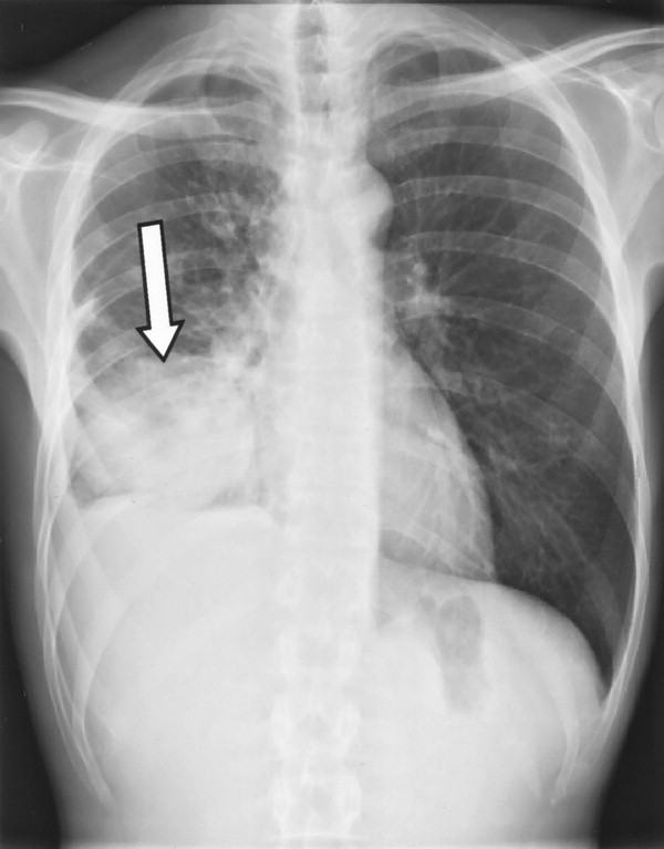
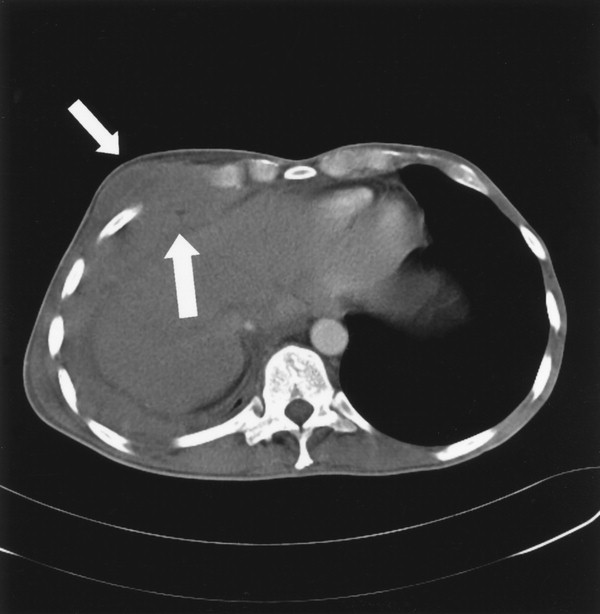
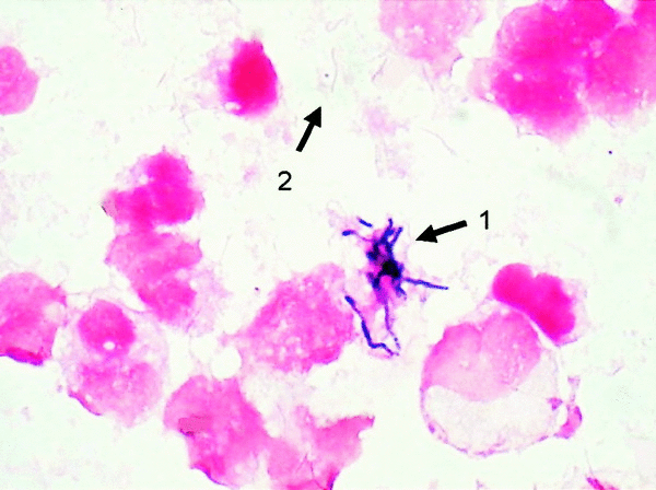
A 43-YOM had cachexia, productive cough and diffuse chest pain ชายอายุ 43 ปีถูกส่งต่อมาเพราะมี cachexia, productive cough, diffuse chest pain, และ chest radiograph มีความผิดปกติที่ปอดข้างขวา (รูป 1). ผู้ป่วยมีประวัติติดสารเสพติดแต่ไม่เคยป่วยเป็นโรคใดๆ. ที่โรงพยาบาลใกล้บ้าน แพทย์ส่งตรวจหาเชื้อวัณโรค แต่ก็ยังไม่พบ. On physical examination, the patient appeared to be moderately ill, with generalized wasting of body muscle and subcutaneous fat. Body weight was 48 kg (body mass index, 16.1). He had a low-grade fever and tachycardia. The oral cavity showed dental caries. There was no local or generalized lymphadenopathy. A slightly painful swelling was palpable in the right sixth intercostal space. Percussion of the right hemithorax was dull and tender. Diminished breathing sounds were heard on auscultation, without crackles or rales. The abdomen was not painful, and there was no organomegaly. Lab: CBC: WBC 26 900/L, Hb level, 5.2 mmol/L), platelet count, 819 000/L), and an elevated C-reactive protein level (200 mg/L). Liver enzyme levels and the results of kidney function tests were normal. Bronchoscopy ไม่พบ endobronchial abnormalities, และการตรวจ bronchoalveolar lavage fluid และ transbronchial lung biopsy samples ไม่ช่วยให้การวินิจฉัย Posted by : cpantip , E-mail : (chpantip@medicine.psu.ac.th) , Date : 2010-04-09 , Time : 15:51:10 , From IP : 172.29.3.68 |

