ความคิดเห็นทั้งหมด : 2

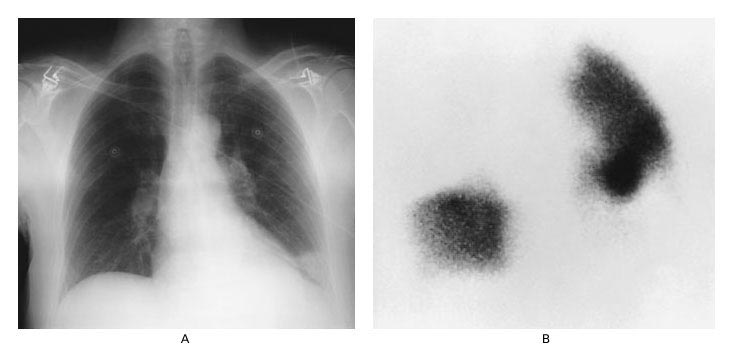
A 67-YOM had cough & progressive dyspnea for 5 days and chest pain for 1 day


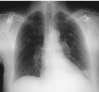
   ชายอายุ 67 ปีมาตรวจที่แผนกฉุกเฉินเนื่องจากมีอาการไอแห้งๆ และหอบเหนื่อยซึ่งเป็นมากขึ้นมา 5 วัน และเจ็บหน้าอกข้างขวาเวลาหายใจลึกๆหรือไอมา 1 วัน. 4 วันก่อน ผู้ป่วยได้รับการวินิจฉัยว่าเป็น left-lower-lobe pneumonia และได้รับการรักษาด้วยยาปฏิชีวนะรูปยากิน.
On examination, the patient had marked respiratory difficulty, with a respiratory rate of 28/min. His lung fields were clear to auscultation.

An arterial blood gas while breathing room air: pH 7.46, PCO2 34 mm Hg, and PO2 61 mm Hg.
ในรูป A คือ CXR ของผู้ป่วย

1. การวินิจฉัยโรคที่น่าจะเป็นมากที่สุดคืออะไร
2. จะ manage อย่างไรต่อ.


Posted by : cpantip , E-mail : (chpantip@medicine.psu.ac.th) ,
Date : 2010-04-08 , Time : 11:51:41 , From IP : 172.29.3.68


ความคิดเห็นที่ : 1


   ขอตอบค่ะ
ชายอายุ 67 ปี
ปัญหา
1. dry cough and progressive dyspnea 5 วัน และ right pleritic chest pain 1 วัน.
2. has been DX left-lower-lobe pneumonia , not respond to oral antibiotic.
3. marked respiratory difficulty, with a respiratory rate of 28/min and lungs clear.
4. Respiratory alkalosis with severe hypoxia (PO2 61 mm Hg)
5. CXR: haziness in wedge shape (pleural base): pulmonary infarction?? ดูเนื้อปอดข้างขวาดำกว่าข้างซ้ายด้วย.

1. การวินิจฉัยโรคที่น่าจะเป็นมากที่สุดคือ acute pulmonary embolism ค่ะ
2. management:
investigation: ทำ multidetector-row spiral CT scan หรือ ventilation-perfusion lung scan
treatment: heparin IV แล้วเปลี่ยนเป็น warfarin กินเมื่อผู้ป่วยอาการดีขึ้น

การป้องกัน ต้องหาแหล่งของ pulmonary embolism เช่น DVT และภาวะ hypercoagulable state


Posted by : lara , Date : 2010-04-11 , Time : 12:01:11 , From IP : 172.29.3.68

ความคิดเห็นที่ : 2


   คุณหมอ lara เก่งมากค่ะ

A chest radiograph demonstrated a pleural-based, wedge-shaped pulmonary infarction (Hampton”s hump) at the left lung base (รูป A). There was focal avascularity (Westermark”s sign) in the right upper lung field.

A ventilation–perfusion scan showed normal ventilation images and loss of perfusion to the entire right upper lobe as well as to the anterior, lateral, and medial basal segments of the left lower lobe (รูป B). Multiple, small perfusion defects were also evident in the left upper lobe. These findings corresponded with the radiographic findings.

ผู้ป่วยได้รับการวินิจฉัย multiple pulmonary emboli และได้รับการรักษาด้วย heparin infusion นาน 5 วันตามด้วย warfarin therapy นาน 6 เดือน.
จากการติดตามที่ 6 เดือน ผู้ป่วยสบายดี. ในผู้ป่วยรายนี้ ตรวจไม่พบปัจจัยเสี่ยงของ pulmonary embolism.

Reference: Sokolove PE, Offerman SR. Pulmonary embolism. NEJM 2001;345 (18): 1311.


Posted by : cpantip , Date : 2010-04-13 , Time : 15:45:57 , From IP : 172.29.3.68

สงวนสิทธิ์การแสดงความคิดเห็นสำหรับ สมาชิกเท่านั้น