
คุณหมอ lara ตอบได้ถูกต้องค่ะ เก่งมากค่ะ
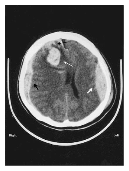
The axial section of a computed tomographic (CT) scan of the head, obtained without the addition of contrast medium, revealed four types of acute post-traumatic intracranial hemorrhage:
1. an epidural hematoma (thick white arrow) and a squamous temporal fracture (which is not shown) on the left side,
2. a laminated subdural hematoma (thick black arrow) on the right side,
3. right-sided periventricular and frontal-lobe contusions containing an intraparenchymal hematoma (thin white arrow), and
4. a subarachnoid hemorrhage (thin black arrow) in the right frontal region.
ทั้งๆที่ได้รับ emergency surgical decompression, ผู้ป่วยเสียชีวิต 5 วันหลังจากการได้รับอุบัติเหตุ.
ตำแหน่งของ acute post-traumatic intracranial hemorrhage, ชนิดของหลอดเลือดที่มี injury และ architecture ของ tissue ล้วนมีผลต่อการปรากฏของ extravascular blood ใน CT scans.
- Epidural hemorrhages มักเกิดจากการฉีกขาดของ meningeal arteries, มักสัมพันธ์กับกะโหลกศีรษะแตก. epidural blood สามารถเซาะ dura จากกะโหลกและทำให้เกิด lenticular hematoma ซึ่งปรากฏเป็น hyperdense ใน CT scanning and that span dural attachments between suture lines.
- Subdural hematomas เกิดขึ้นเมื่อเลือดรั่วออกมาเข้าสู่ subdural space ระหว่าง arachnoid และ pia mater. Subdural hematomas surround the brain, forming crescent-shaped hyperdense areas ใน CT scanning ซึ่งสามารถแทรกเข้าไปใน gyri ของ cortex ไปตาม hemisphere.
- การแตกของ intraparenchymal capillaries ซึ่งมีผลทำให้เกิด contusion ปรากฎเป็น area of intermediate density ใน CT scanning. intraparenchymal hemorrhage ที่ไปแย่งที่เนื้อสมองปรากฏเป็น hyperdense area ใน CT scanning ร่วมกับมี hypodense (edematous) อยู่รอบๆ.
-Trauma เป็นสาเหตุที่พบบ่อยที่สุดของ subarachnoid hemorrhage, ซึ่งปรากฎใน subarachnoid space.
Reference: Mattiello JA, Munz M. Four Types of Acute Post-Traumatic Intracranial Hemorrhage. NEJM 2001;344 (8): 580.
Posted by : cpantip , Date : 2010-04-05 , Time : 09:57:12 , From IP : 172.29.3.68
|